

Superimpose Proteins
The Superimpose Proteins plugin overlays 2 or more proteins in 3D space for visual comparison and calculates root-mean-square deviation (RMSD) values, a number in Å that measures how far apart the structures are.
Protein overlay uses either the backbone (alpha carbons) or all heavy protein atoms. The alignment can use the full protein, one chain, a ligand's binding site, or a selection.
A Fixed Reference structure is chosen, and Moving Structures are rotated and moved onto the Fixed Reference. RMSD values are reported between the Fixed Reference and each Moving Structure.
Instructions
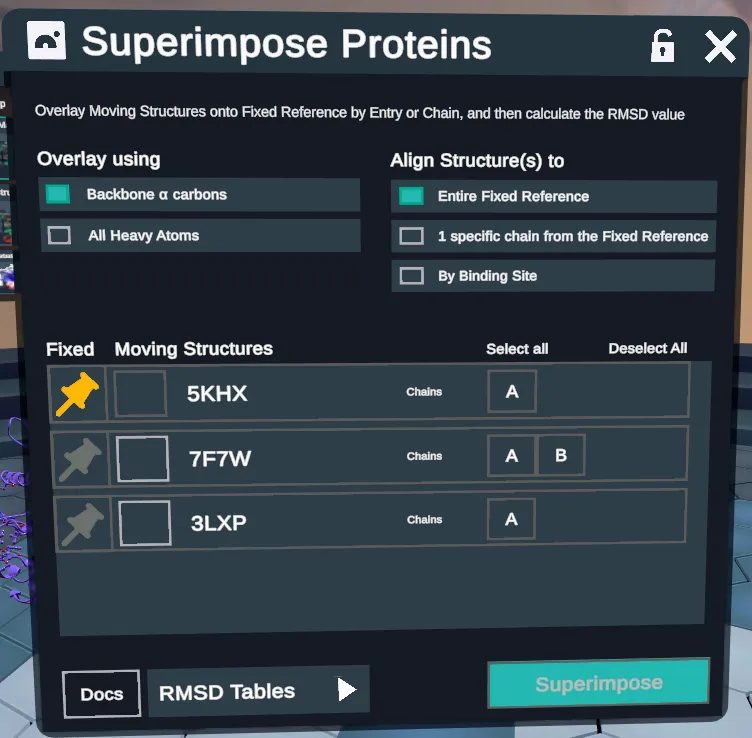
- In the upper left, under Overlay using, pick how structures are overlaid.
- Backbone α carbons: Overlay structures based on all paired alpha carbons between the 2 proteins.
- All Heavy Atoms: Uses backbone atoms and side chain atoms (all protein atoms except for hydrogen) for paired residues.
- In the upper right, under Align Structure(s) to, there are 4 alignment modes.
- Entire Fixed Reference: Uses a sequence based alignment to superimpose entire proteins on top of each other.
- 1 specific chain from the Fixed Reference: Uses a sequence based alignment to superimpose the moving structures onto a specified chain on the Fixed Reference.
- By Binding Site: The user picks a ligand on the Fixed Reference, and each Moving Structure is aligned onto the binding site around it, using Site-Motif (see Methodologies).
- By Selection (not in this screenshot; see step 6): The user selects specific residues in the Fixed Reference and the moving structures are aligned by that selection only. Users may choose to specify residues on both the fixed and moving structures but only selections in the Fixed Reference are required for the plugin to run.
- A list of proteins in the workspace appears in the main panel. In this panel, choose Fixed Reference and Moving Structures, and pick a chain for each when aligning by chain, or a ligand on the Fixed Reference when aligning by binding site (see 4 and 5, below).
- By default, the first entry is set as Fixed, and designated by the yellow pin. When selected as Fixed, the box under Moving will be inactive. To change this selection, select the pin next to the desired Fixed Reference protein.
- Choose one or more Moving Structures by selecting the boxes under the Moving Structures column. This box will turn green when selected.
- Once selections are made, the Superimpose button in the lower right will become active and show the number of proteins selected. (e.g. “Superimpose (3)” for a Fixed Reference and 2 Moving Structures). Click this button to superimpose the proteins and calculate RMSD values. The button will change to “Calculating...” as the process is being completed.
- Once complete, the proteins will be superimposed and locked, for visual inspection. The RMSD tables pulldown menu in the lower left will become active. Each superimpose run will generate a new table, with the most recent run appearing at the top of the pulldown list. Select the run to open the table in the workspace. Tables can be closed and reopened in the workspace.
- Overlays all paired alpha carbons. Picked here.
- Overlays backbone and side chain atoms, all but hydrogen.
- Sequence alignment of the entire proteins. Picked here.
- Aligns the moving structures onto one chain of the Fixed Reference.
- Aligns each Moving Structure onto the binding site around a ligand on the Fixed Reference, using Site-Motif.
- The yellow pin marks the Fixed Reference. Select another pin to change it.
- Select one or more. A selected box turns green.
- The chain for each protein, when aligning by chain.
- One table per run, newest at the top. Active after a run.
- Runs the overlay and RMSD. Active once selections are made.
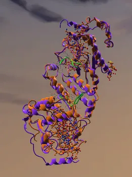
2 superimposed proteins
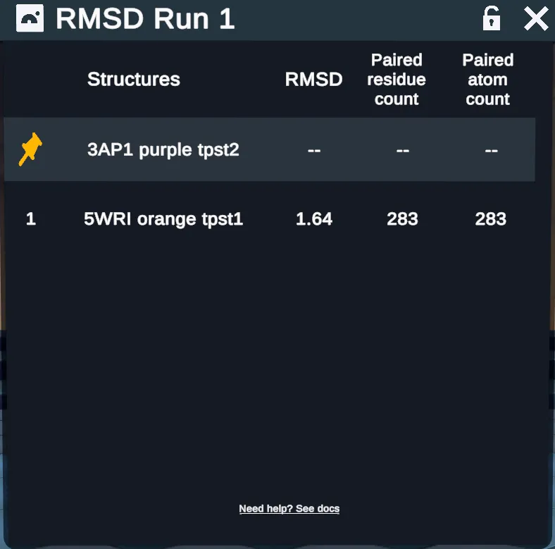
RMSD table describing above superimposed proteins
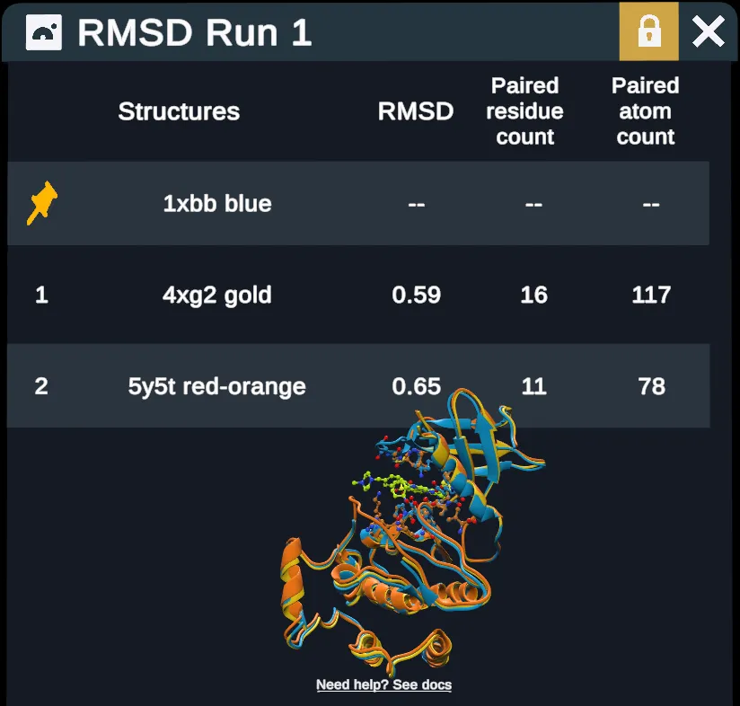
- The top row, marked with the yellow pin.
- One numbered row each.
- Values close to zero mean highly similar proteins.
- Residues paired between the 2 structures.
- Atoms paired. Equals the residue count for backbone α carbons.
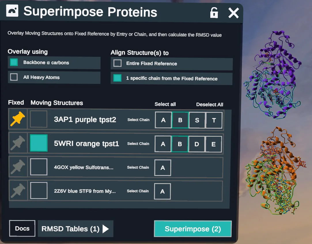
- When aligning by chain, the main panel chain selection boxes will become active. Choose Fixed and Moving Structures as above, and additionally choose a chain for each protein to be superimposed
- Note that selecting a chain on the plugin main panel will also apply green highlighting to the selected chain of the protein structure, to visualize the selections before running.
- Picked here.
- 3AP1 purple tpst2 is the Fixed Reference.
- 5WRI orange tpst1 is selected.
- The chain for each protein. Chain B is picked on 3AP1 and 5WRI.
- The picked chains show green on the structures.
- The Fixed Reference and one Moving Structure.
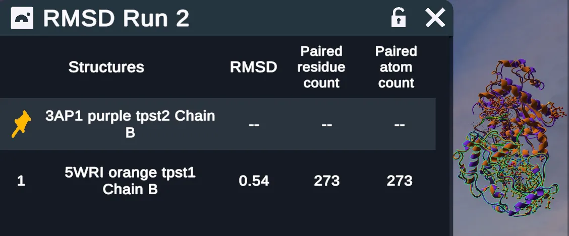
The RMSD table for a chain run names the chain with each structure.
- 3AP1 purple tpst2, chain B.
- 5WRI orange tpst1, chain B, at RMSD 0.54.
- The 2 proteins after the run.
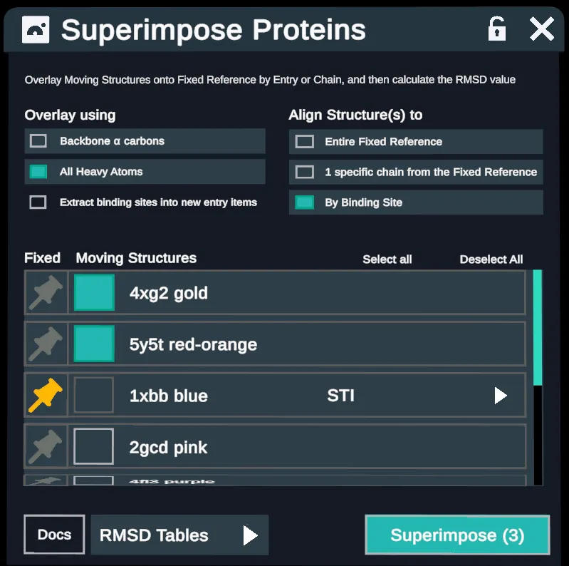
When aligning by Binding Site, the Overlay method will automatically change to All Heavy Atoms.
- An option to Extract binding sites into new entry items appears. This creates a new set of entries containing only the superimposed binding site atoms of each protein.
- Choose Fixed and Moving Structures as above and additionally choose a ligand from the Fixed Reference to establish the binding site for superimposition.
Note the Extract binding sites into new entry items button
- Picked here.
- Picked automatically.
- New entries with only the superimposed binding site atoms.
- 1xbb blue is the Fixed Reference.
- The ligand that sets the binding site. STI here.
- 4xg2 gold and 5y5t red-orange are selected.
- The Fixed Reference and 2 Moving Structures.
When aligning by selection, the user must make a selection in the Fixed Reference. These selections can be made by using the selector tool on the 3D structure itself, or by using menus such as the Entry Hierarchy and the Antibody Regions table.
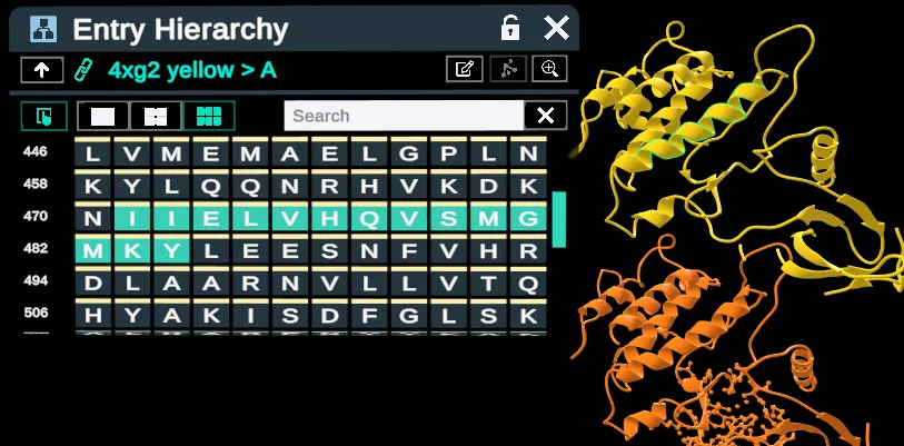
Making a selection in the Fixed Reference using the Entry Hierarchy.
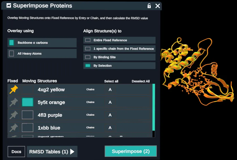
Structures superimposed based on a selection in the Fixed Reference
RMSD tables report out fixed and moving structures (with chain when relevant) RMSD values, paired residues, and paired atoms
- Note that when superimposing by backbone alpha carbons, the # of paired residues = # of paired atoms; when superimposing by all heavy atoms, the # of paired residues will be ~8x less than the # of paired atoms because there are on average ~8 heavy atoms per residue.
- For 2 identical proteins, the RMSD would be zero. Values close to zero indicate highly similar proteins. Values over 10 Å mean very different structures. A combination of visual inspection and RMSD values gives a reasonable indication of how similar the structures are.
Methodologies
We have 2 main approaches to superimposing proteins:
Full protein, chain and selection alignments use a sequence based approach.
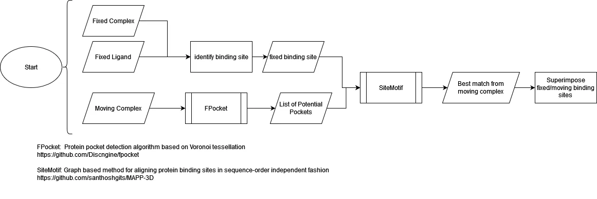
Binding site overlays use the Site-Motif algorithm, with fpocket for identifying inputs.
Sequence Alignment
Primary modules
Bio.pairwise2
Bio.PDB.Superimposer
Summary
For each pairing of fixed structure and moving structure:
A global alignment is run on protein residue sequences using Bio.pairwise2
Aligned residue atoms are passed into Bio.PDB.Superimposer, which calculates an RMSD value and a transform matrix.
We apply the transform matrix to every moving atom on the complex.
After all moving atoms have been transformed, we update the complexes in the workspace.
Implementation Details
Alignment
- For Entire Fixed Reference, the whole protein sequence is aligned.
- For 1 specific chain, only the chosen chains are aligned.
- For By Selection, only the selected residues are aligned. A Moving Structure with no selection is aligned whole.
Global alignment scoring values
- match = 2
- mismatch = -1
- gap open penalty = -10
- gap extend penalty = -.5
Superimpose
- If the alpha carbon overlay method is selected, we only pair the alpha carbons from each residue.
- If all heavy atoms overlay method is selected, we attempt to pair all heavy atoms. In some cases, we cannot get a 1-1 pairing of heavy atoms. In this case, those residues are excluded from the superimpose
Binding Site Alignments using Site-Motif
Site-Motif is described as a graph based method for aligning protein binding sites in sequence-order independent fashion. It can analyze the binding site of a protein and find the best alignment on another protein.
Our algorithm uses fpocket to identify potential binding sites on the moving protein, and those results are compared to the fixed binding site using Site-Motif. Site-Motif calculates alignments of each fpocket result to the fixed binding site, and the result with the longest alignment is used as the corresponding binding site. A transformation matrix is calculated to superimpose the moving binding site onto the fixed.
Source Code
Note that Nanome maintains a fork of the code from the original paper. The original code is MAPP-3D on GitHub.
Improvements to the codebase include:
- Ported to Python 3
- Removed features we don't use
- Added logging
Overview
References
Sankar S, Chandra N (2022) SiteMotif: A graph-based algorithm for deriving structural motifs in Protein Ligand binding sites. PLoS Comput Biol 18(2): e1009901. https://doi.org/10.1371/journal.pcbi.1009901
Le Guilloux, V., Schmidtke, P. & Tuffery, P. Fpocket: An open source platform for ligand pocket detection. BMC Bioinformatics 10, 168 (2009). https://doi.org/10.1186/1471-2105-10-168