

Main Menus
Entry List
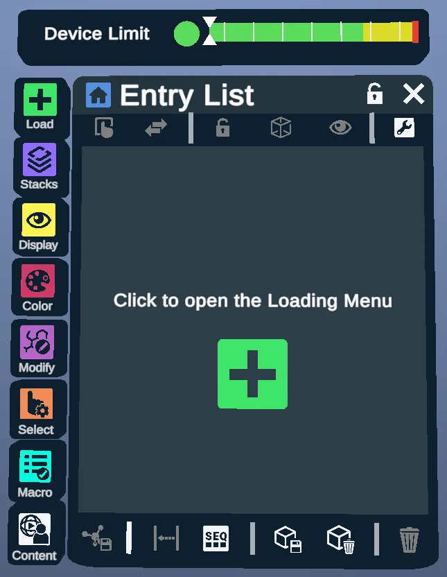
The Entry List is a list of all of the structures that have been loaded into the environment.
The Load, Stacks (plugins), Display, Color, Modify, Select, Macro, and Content (spatial tutorials) Menus can be displayed by clicking their respective icons down the left side of the Entry List.
Device Limiter
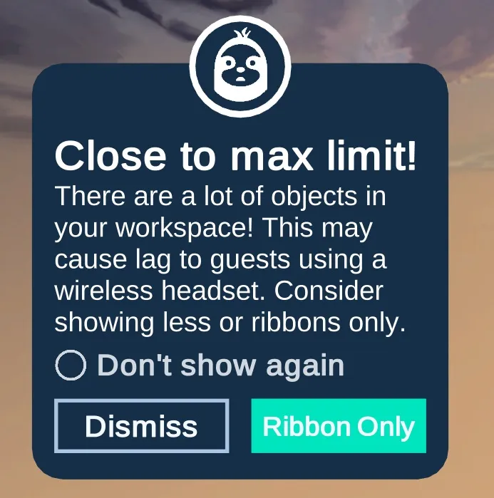
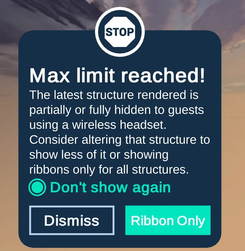
The Device Limit bar compares how much your workspace shows, atoms and ribbons alike, with what all-in-one (wireless) headsets can draw. When it gets close to the limit or reaches it, one of these prompts appears. Each warns that guests on a wireless headset may see lag or hidden structures, and offers Dismiss or Ribbon Only, plus a Don't show again box.
Hierarchy Menu
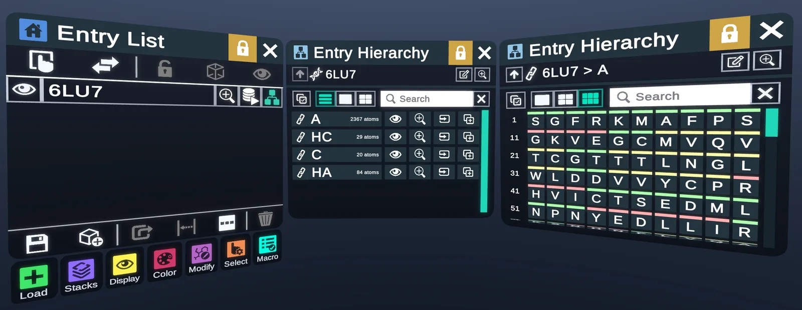
- HierarchyOpens the Hierarchy Menu for the structure.
- Frame MenuOpens the Frame Menu for the structure.
- Chains6LU7 at its top level, listing its chains.
- Chain A of 6LU7, listing its residues.
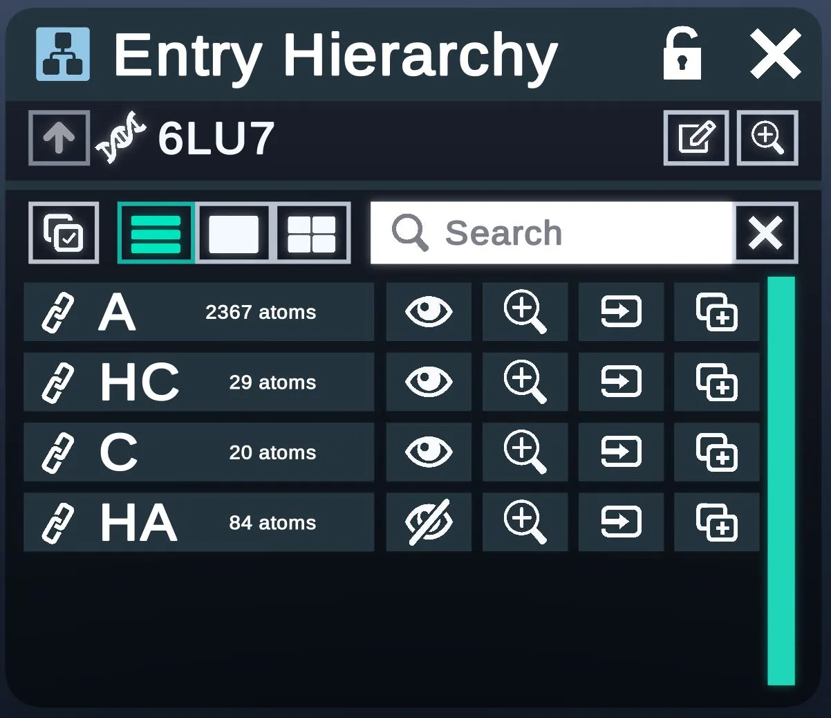
The Hierarchy Menu lists out the components of an entity, such as chains and residues, in hierarchical levels.
- UpGoes back up one level. It's grayed out at the top level, as here.
- The level you're on, such as 6LU7 or 6LU7 > A.
- View modesA list, or icons in different sizes. The list is on here.
- ChainsOne row per chain, with its atom count.
- Show / HideShows or hides each chain. HA is hidden here.
- ZoomZooms to each chain.
Different Viewing Modes (Small, Medium, Large)

Different Viewing Modes can be toggled to modify the substructure icon sizes in each menu.
- Large icons.
- MediumMedium icons.
- SmallSmall icons, as here: one letter per residue.
- Residue numberThe number of the first residue in each row.
| Medium | Large |
|---|---|
 |  |
Frame Menu
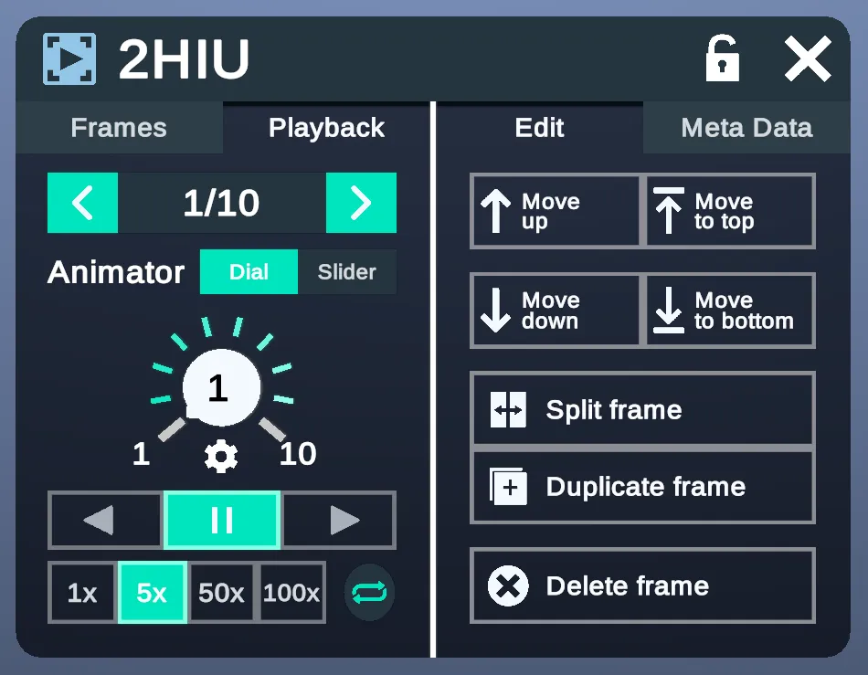
The Frame Menu lets users navigate and modify different frames of the same structure (Academic and Enterprise only).
- FramesThe list of frames, shown below.
- PlaybackShown here. Steps through and plays the frames.
- EditShown here. Moves, splits, duplicates and deletes frames.
- Meta DataThe frame's meta data, shown below.
- Previous / Next frameThe arrows change frame. The middle shows the frame and the total, here 1/10.
- Moves through the frames with a dial or a slider.
- The dial, from the first frame to the last.
- Play / PausePause in the middle, with a play button on each side.
- 1x, 5x, 50x or 100x.
- Loops the playback.
- Move upMoves the current frame up one place.
- Moves the current frame to the top.
- Moves the current frame down one place.
- Moves the current frame to the bottom.
- Splits the current frame.
- Duplicate frameDuplicates the current frame.
- Delete frameDeletes the current frame.
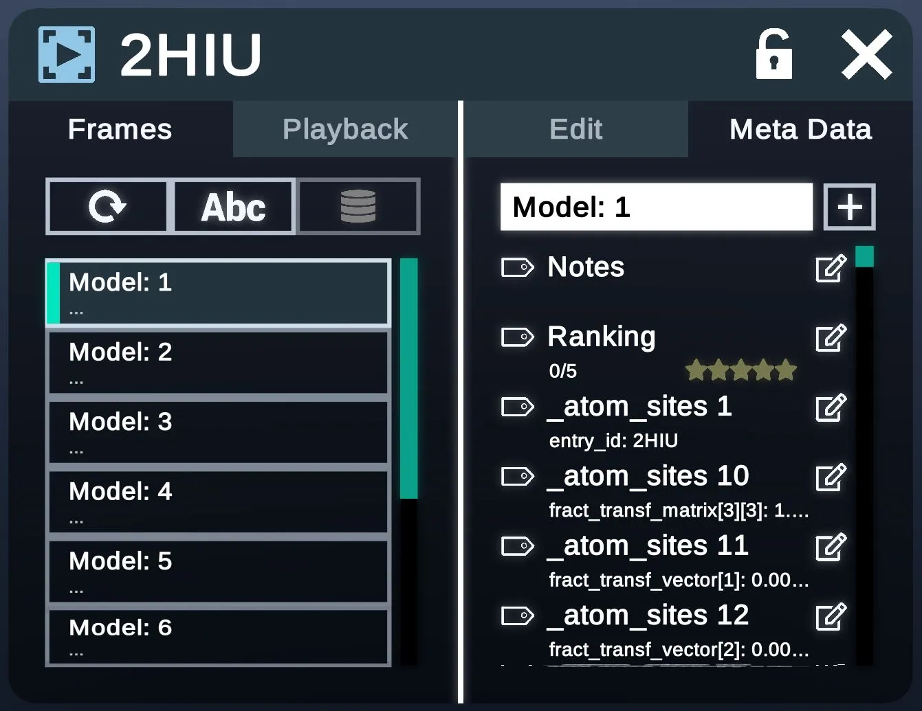
- Model listOne row per frame. Selecting one switches to it.
- Meta dataThe current frame's meta data, such as Notes and Ranking, each with an edit button.
Electron Density Maps
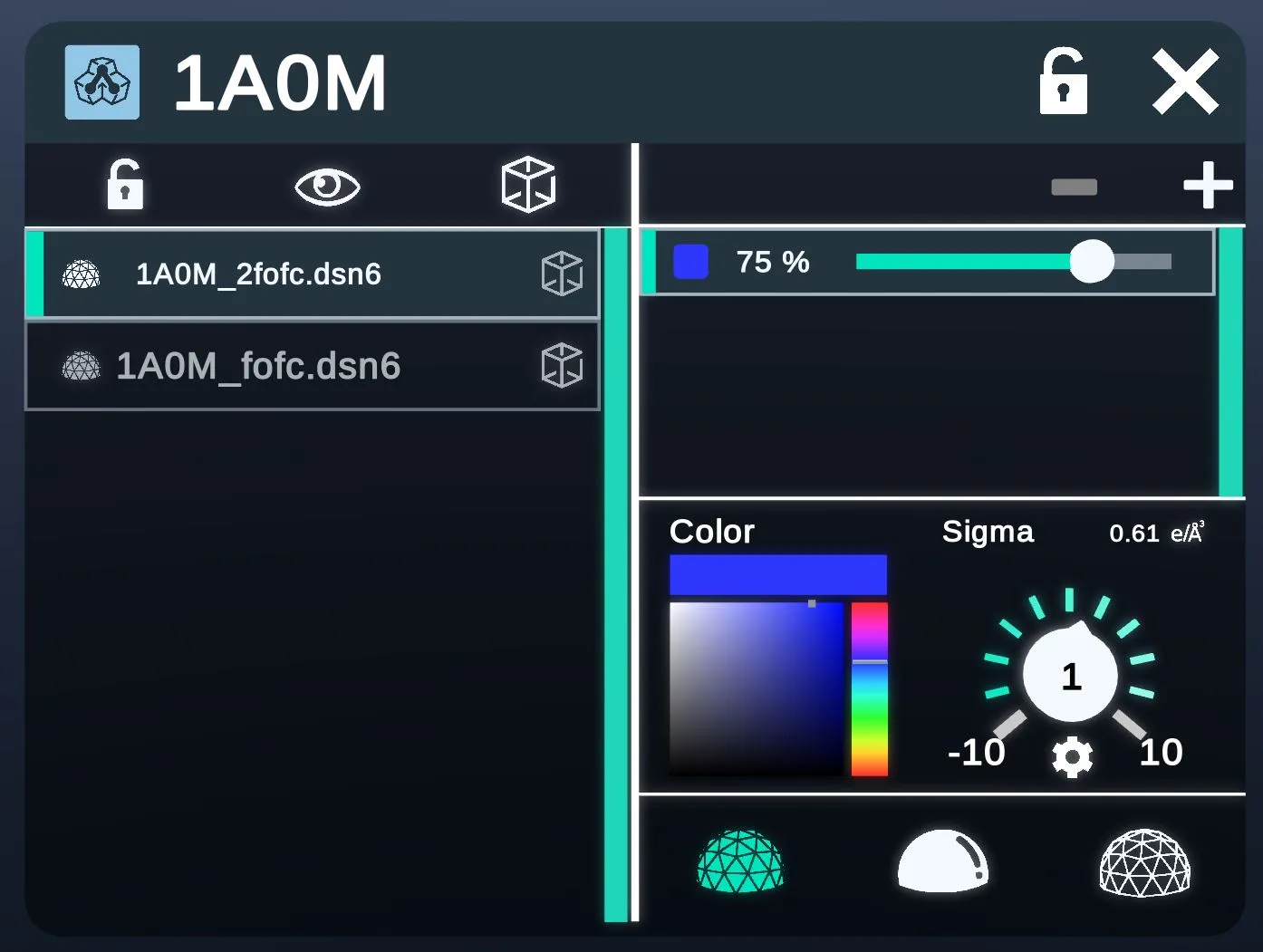
The Electron Density tab shows the configuration of viewing the Electron Density Map of the structure (Academic and Enterprise only).
- MapsThe maps loaded for the structure, here 2 .dsn6 files.
- The map's opacity, here 75 %.
- ColorThe map's color.
- The sigma level, from -10 to 10. Its value in e/ų shows beside it, here 0.61.
- 3 ways to draw the map.
Alternative Conformations Menu
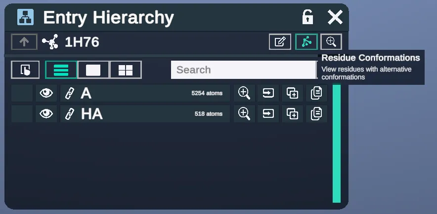
- View residues with alternative conformations. Opens the menu below.
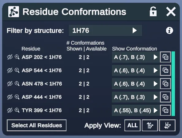
This menu specifies residue conformations.
- Filter by structureLists the residues of one structure, here 1H76.
- Each residue with alternative conformations, with buttons to hide it and zoom to it.
- How many conformations are shown, and how many are available.
- The conformations shown, such as A (.7), B (.3).
- Select All ResiduesSelects all the residues.
- Apply ViewALL and 2 other views.
Menu Actions
Action buttons on the Entry List provide quick access to common actions on the workspace.
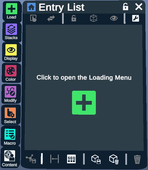
- Select/Deselect AllSelect or deselect all present structures.
- Invert SelectionInvert current selection.
- Toggle LockLock or unlock selected entities.
- Toggle BoxShow or hide the box frame of selected entities.
- Toggle VisibilityShow or hide the selected entities.
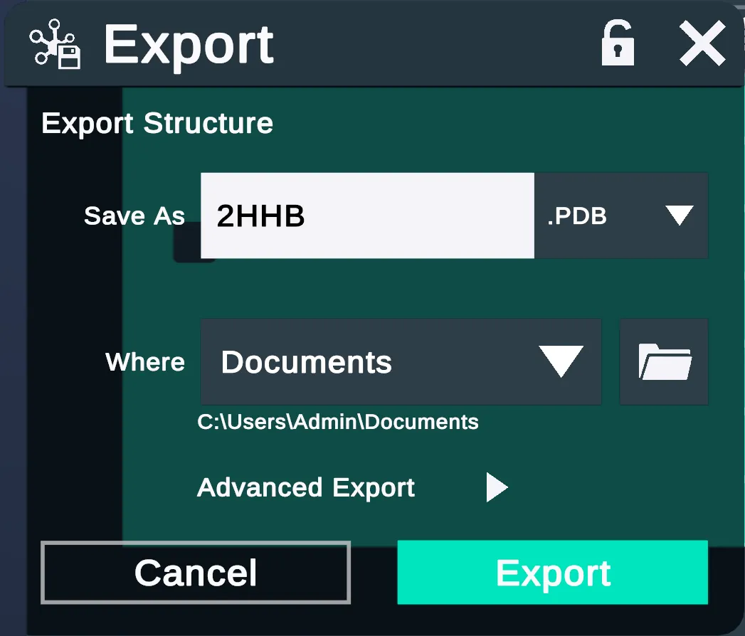
- Export the selected structures in a desired format. Opens the Export menu.
- Align the selected entities and place them in the same position under the same coordinate system. Caution: this does not necessarily align the entities by their atom position, and the entities could end up not overlapping after the alignment.
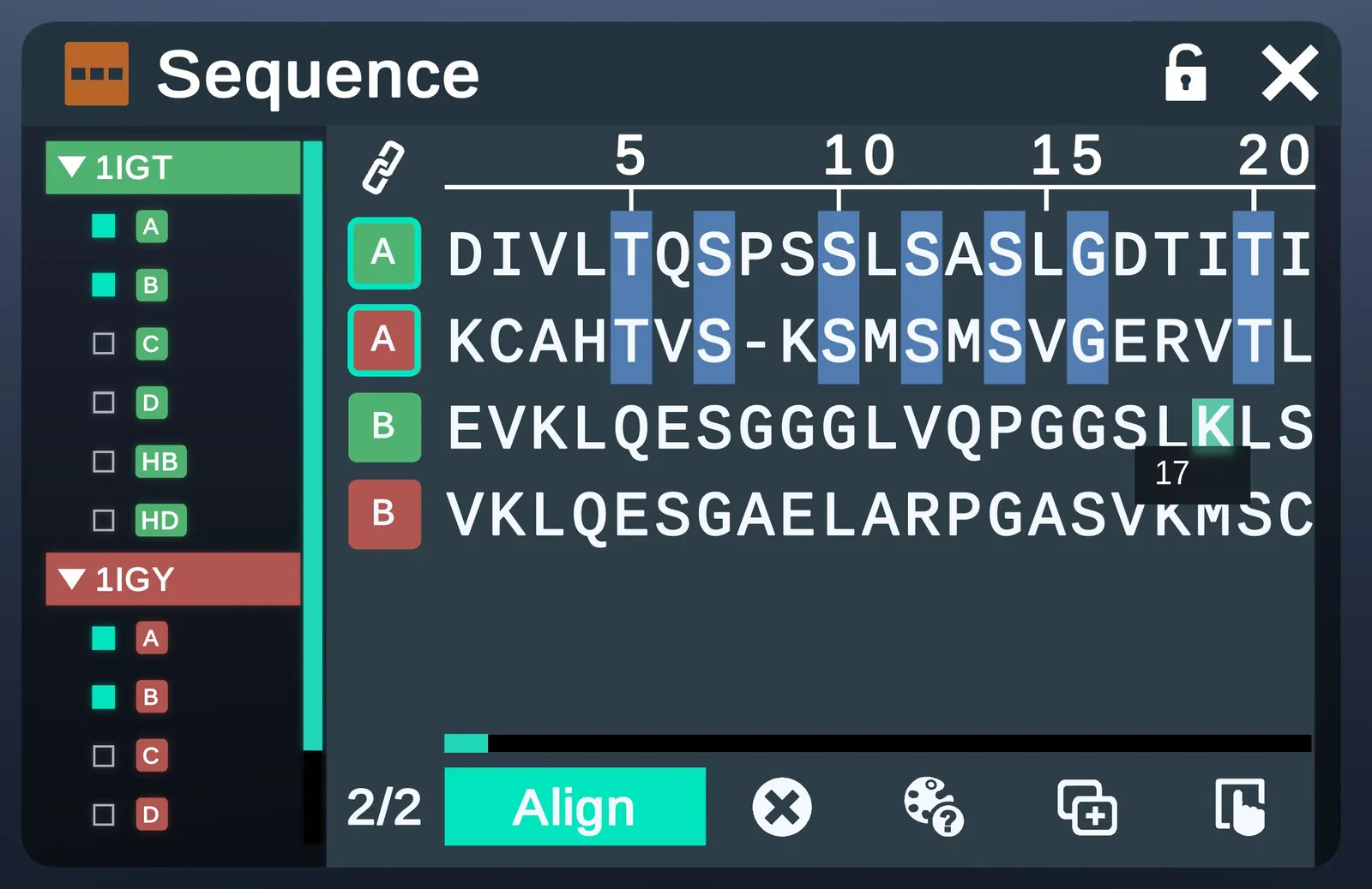
- View the sequences of multiple chains and align them. Opens the Sequence menu.
- SaveSave the current workspace.
- Discard the current workspace and create a new one.
- DeleteDelete the selected entities.
| Export menu | Sequence menu |
|---|---|
 |  |
Spatial ("Quick") Anchor
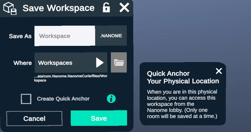
When saving a workspace, you have the option to create a spatial anchor for your current physical location. When you re-launch Nanome in this same physical location, you'll be able to quickly access this workspace from the Nanome Lobby. (Only one workspace can be saved with a spatial anchor at a time.)
Load
The Load menu loads molecules or structures into your workspace. Supported file formats (v1.24):
Structural Coordinate File Formats
.pdb, .cif, .mmcif, .pse, .nanome, .sdf, .mol, .xyz, .mol2, .pqr, .mae (Maestro), .moe (MOE), and SMILES files
Electron Density Map File Formats
.ccp4, .dsn6
Trajectory/Molecular Dynamics File Formats
.dcd, .gro, .trr, .xtc, .psf
Image File Formats
.png, .jpg, .jpeg, .pdf
Gzip-compressed copies (.gz) of supported files load too.
For the file formats supported in Nanome v2, see Supported File Formats.
Featured
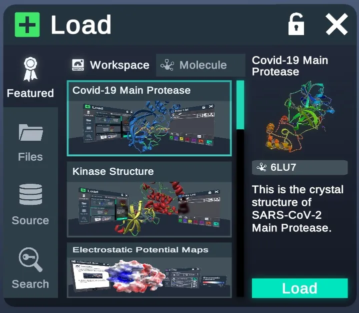
Workspaces that contain featured structures and tutorials.
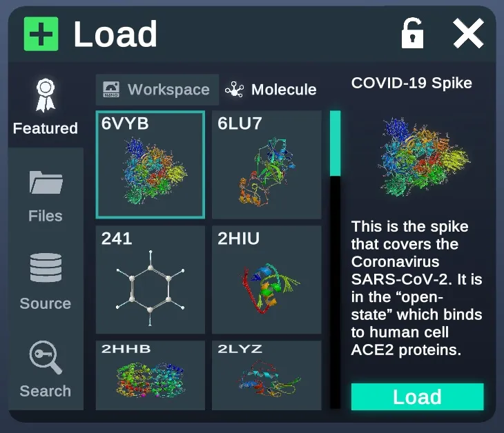
Molecules featured by Nanome for their interesting properties or background.
Files
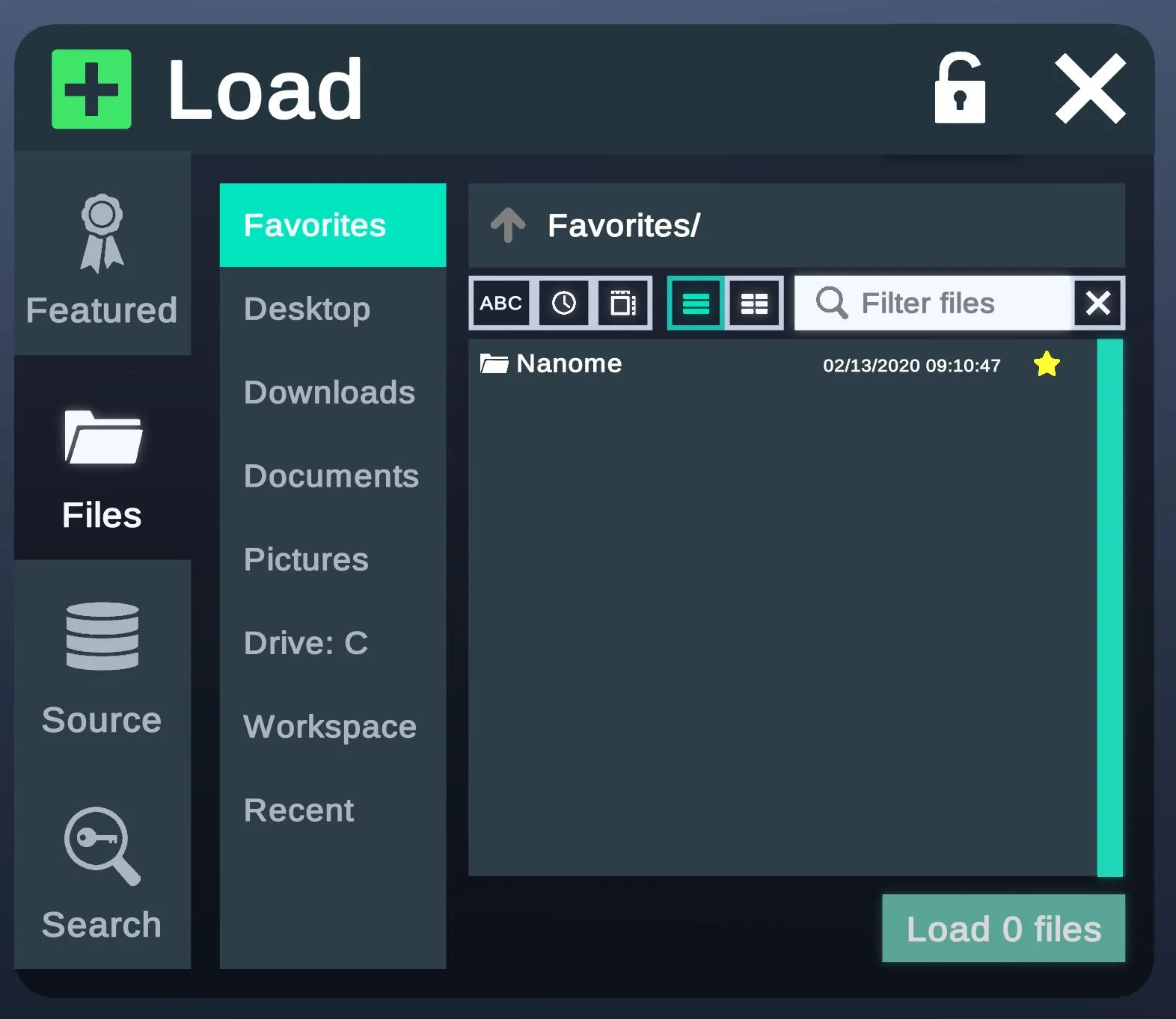
The Files tab loads structures from your computer. Recent files are listed under Files.
Source
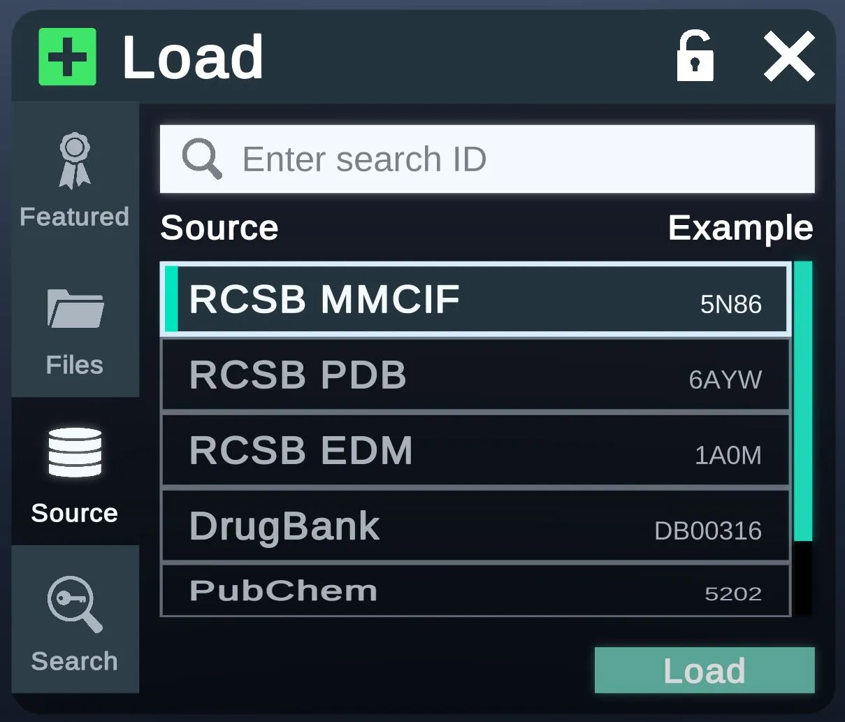
From the Source tab, structures can be loaded directly from RCSB, DrugBank (Academic and Enterprise only) or PubChem (Academic and Enterprise only). A custom database can be added in the config file.
Search
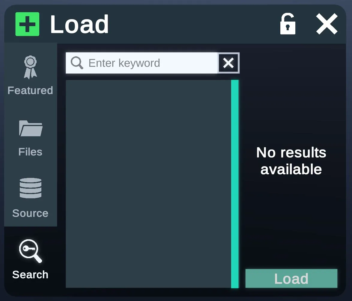
From the Search tab, find and load a structure by keyword.
Stacks
The Stacks tab loads structures from custom databases added through plugins.
Cancel a load
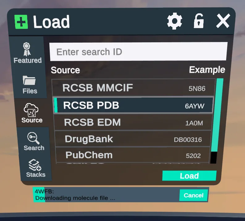 | 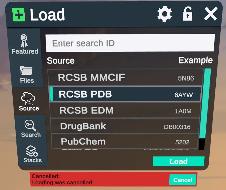 |
Cancel a load from the Source tab if it's taking too long or the PDB code is wrong.
Stacks
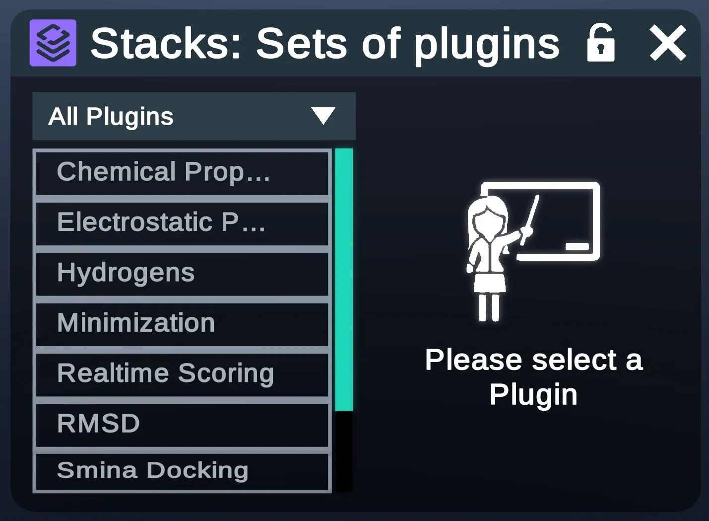
- All PluginsPicks which set of plugins to list.
- Plugin listThe plugins in the set.
- Shows the plugin you select.
The Nanome Stacks page has more.
Display
Atom
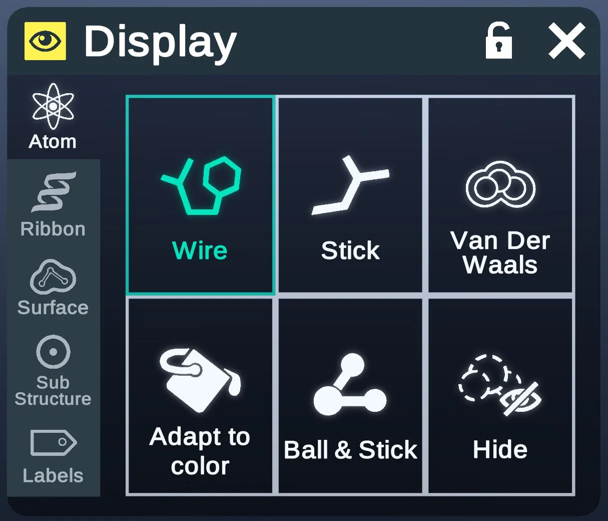
This tab contains options for the visual of atoms and bonds.
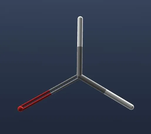
- Hide atoms and show bonds in thin wire visual.
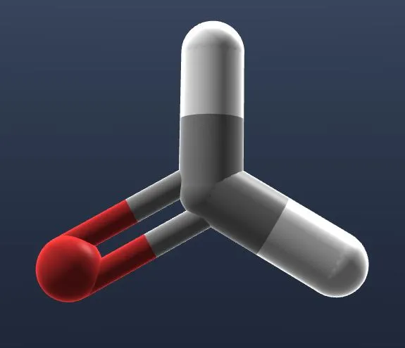
- Show small atoms and bonds in volumetric stick visual.
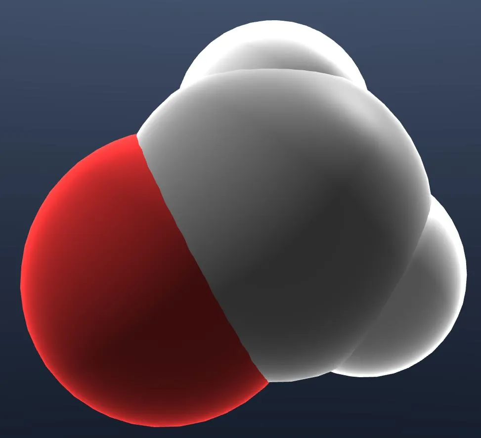
- Shows each atom as a sphere at its van der Waals radius, with no bonds.
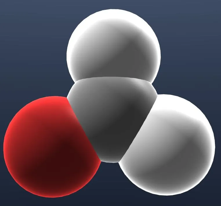
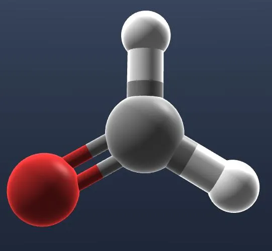
- Also called Adaptive. Shows each atom as a sphere sized by its color, with no bonds.
- Show atoms as small balls joined by stick bonds.
- HideHide all atoms and bonds.
| Wire | Stick | Van Der Waals | Adapt to color | Ball & Stick |
|---|---|---|---|---|
 |  |  |  |  |
Ribbon
This tab contains options for the visual of ribbons.
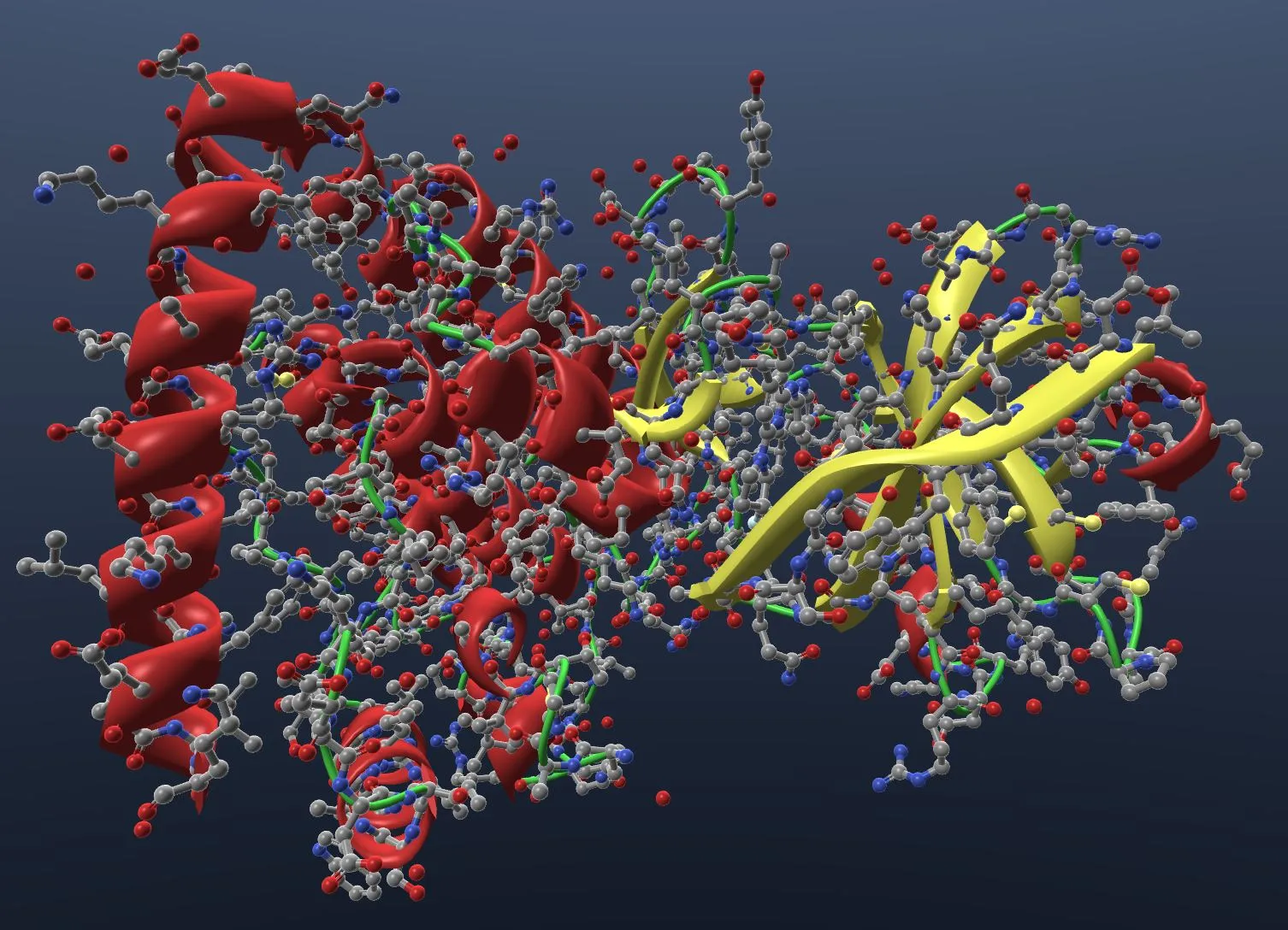
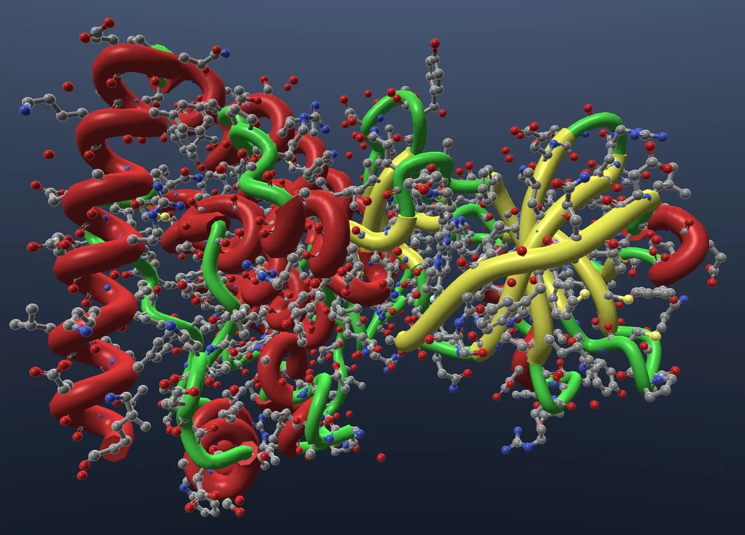
- Shows the ribbon as a cartoon: helices as spirals and strands as arrows.
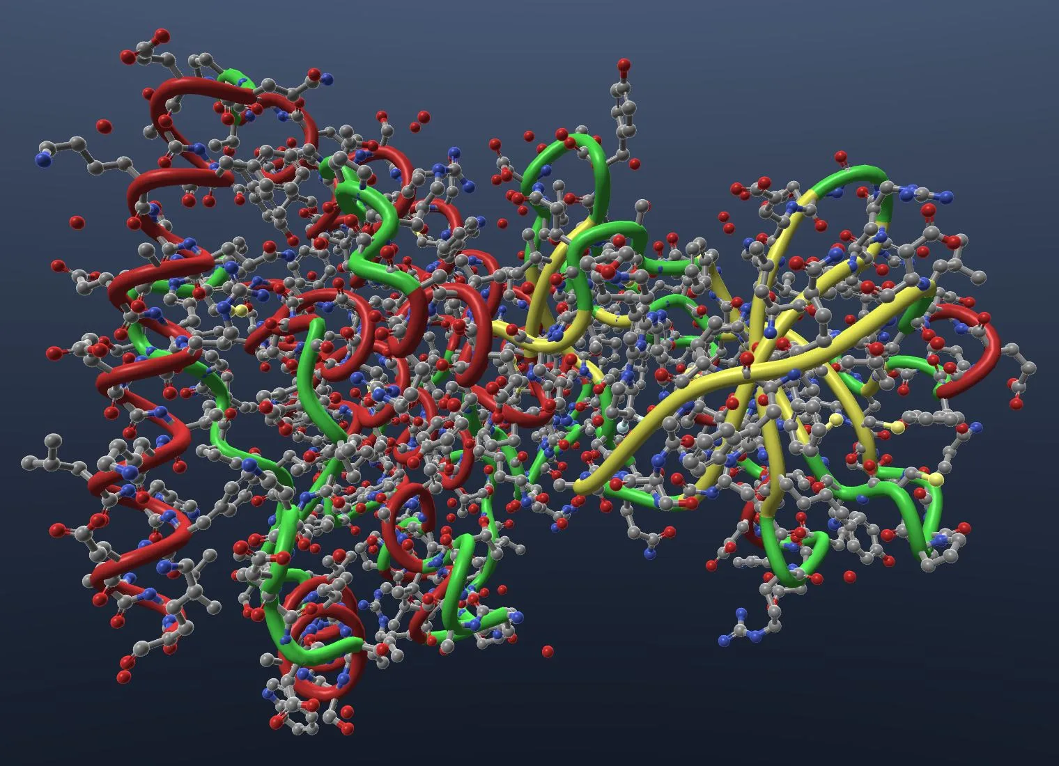
- Show ribbon in uniform tube visual.
- Show ribbon in tube with widths proportional to the atoms' b-factor value.
- HideHide the ribbon.
| Cartoon | Coil | Putty |
|---|---|---|
 |  |  |
Surface
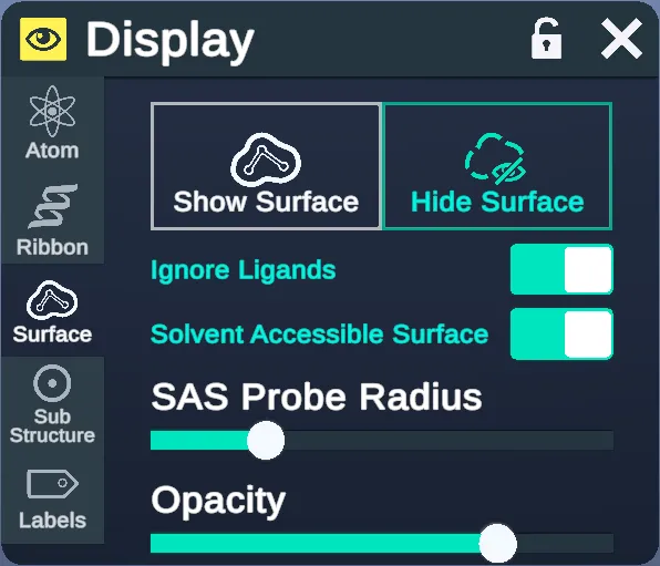
Show or hide the surface, change its opacity, or change the probe radius (in Å).
- Shows the surface.
- Hides the surface.
- Leaves ligands out of the surface.
- Shows the solvent-accessible surface. Off, the surface is solvent-excluded.
- The radius of the solvent probe, in Å.
- The surface's opacity.
Mesh Surfaces (PCVR only)
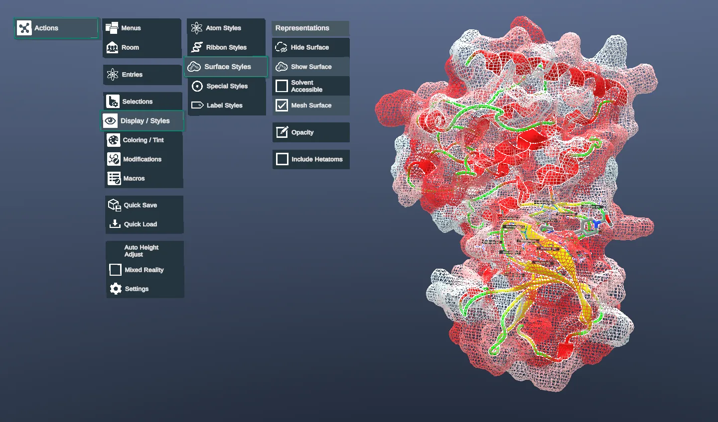
If you are using PCVR, you also have the option to display a mesh surface. This option can be found in the Action Menu. Navigate to Display/Styles > Surface Styles, then check the "Mesh Surface" option.
Sub Structure
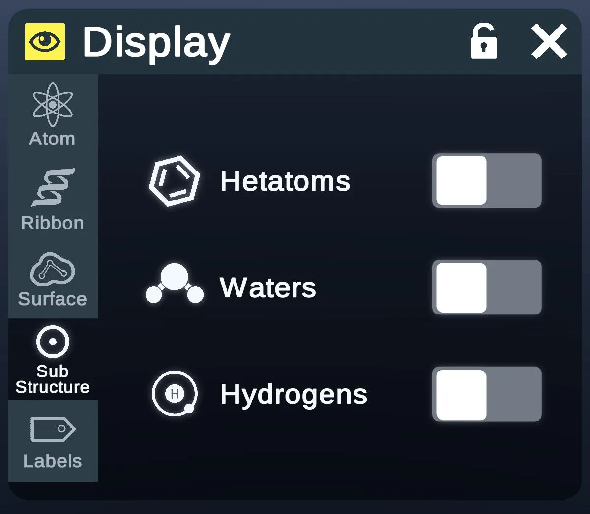
- HetatomsShows hetatoms other than water, such as ligands.
- Shows waters.
- Show hydrogens.
| Hetatoms | Waters | Hydrogens |
|---|---|---|
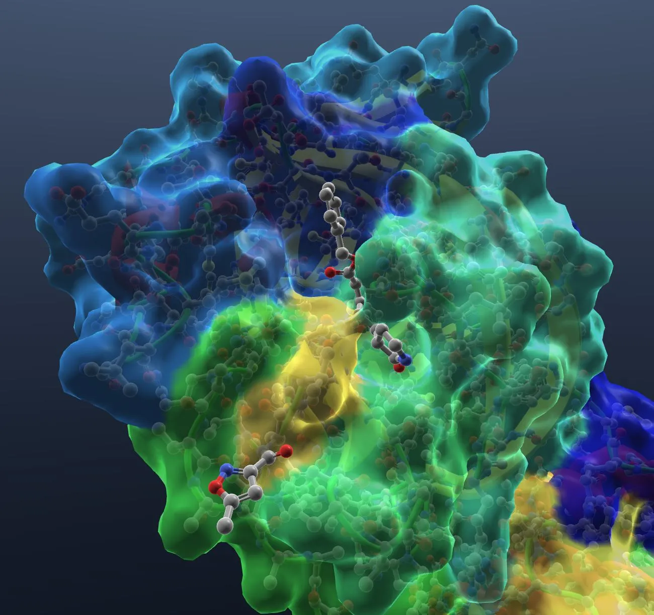 | 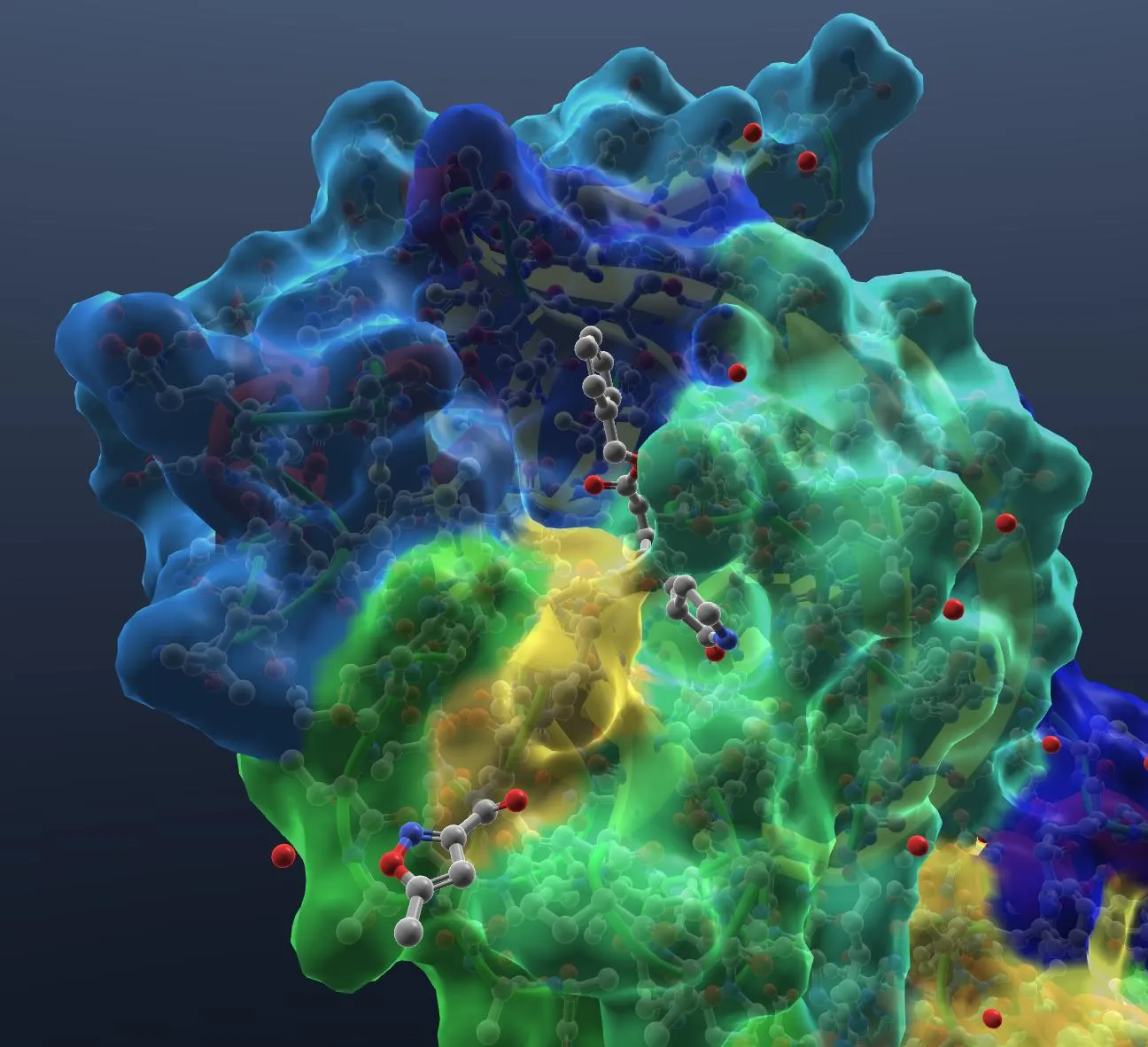 | 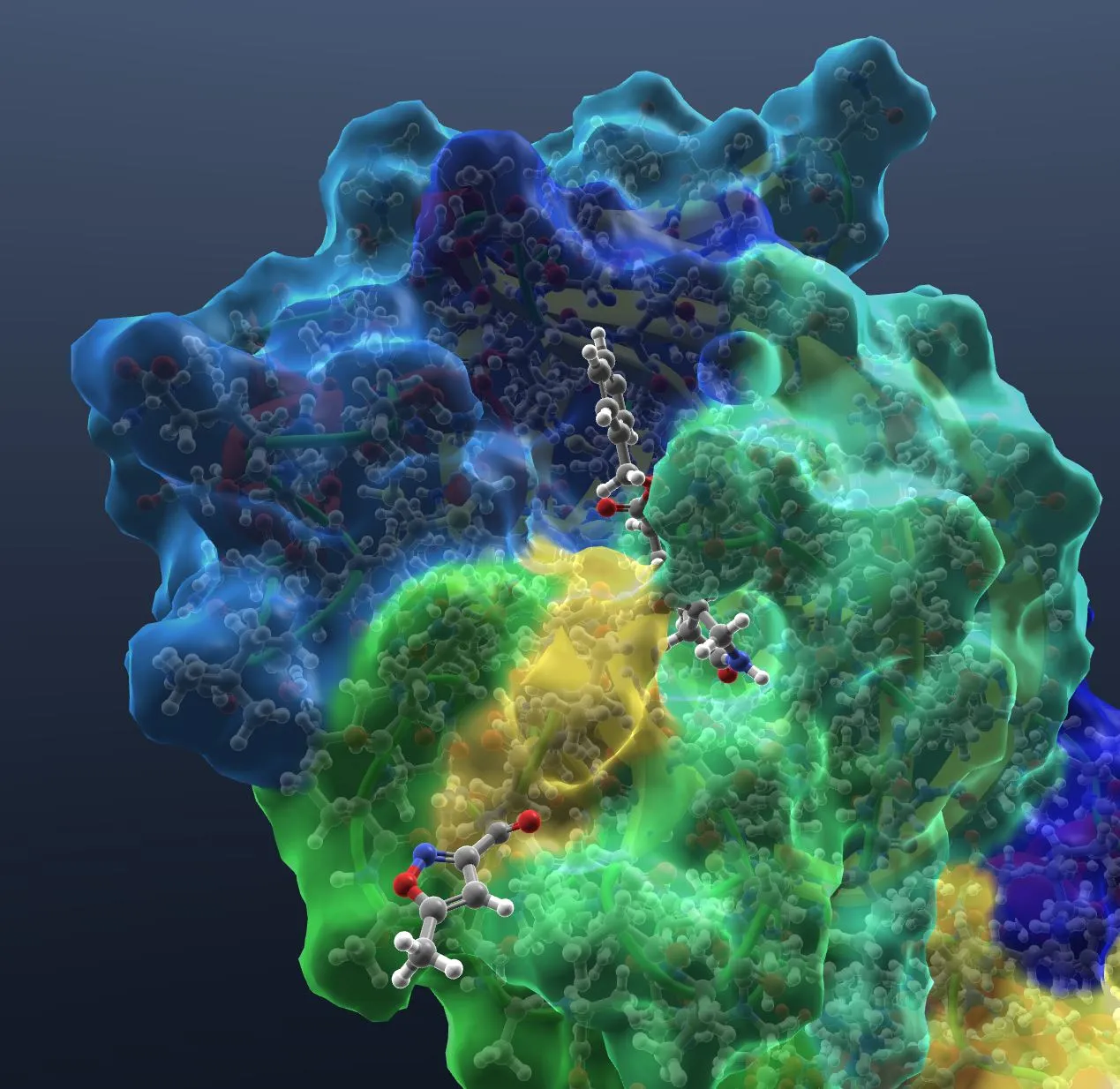 |
Labels
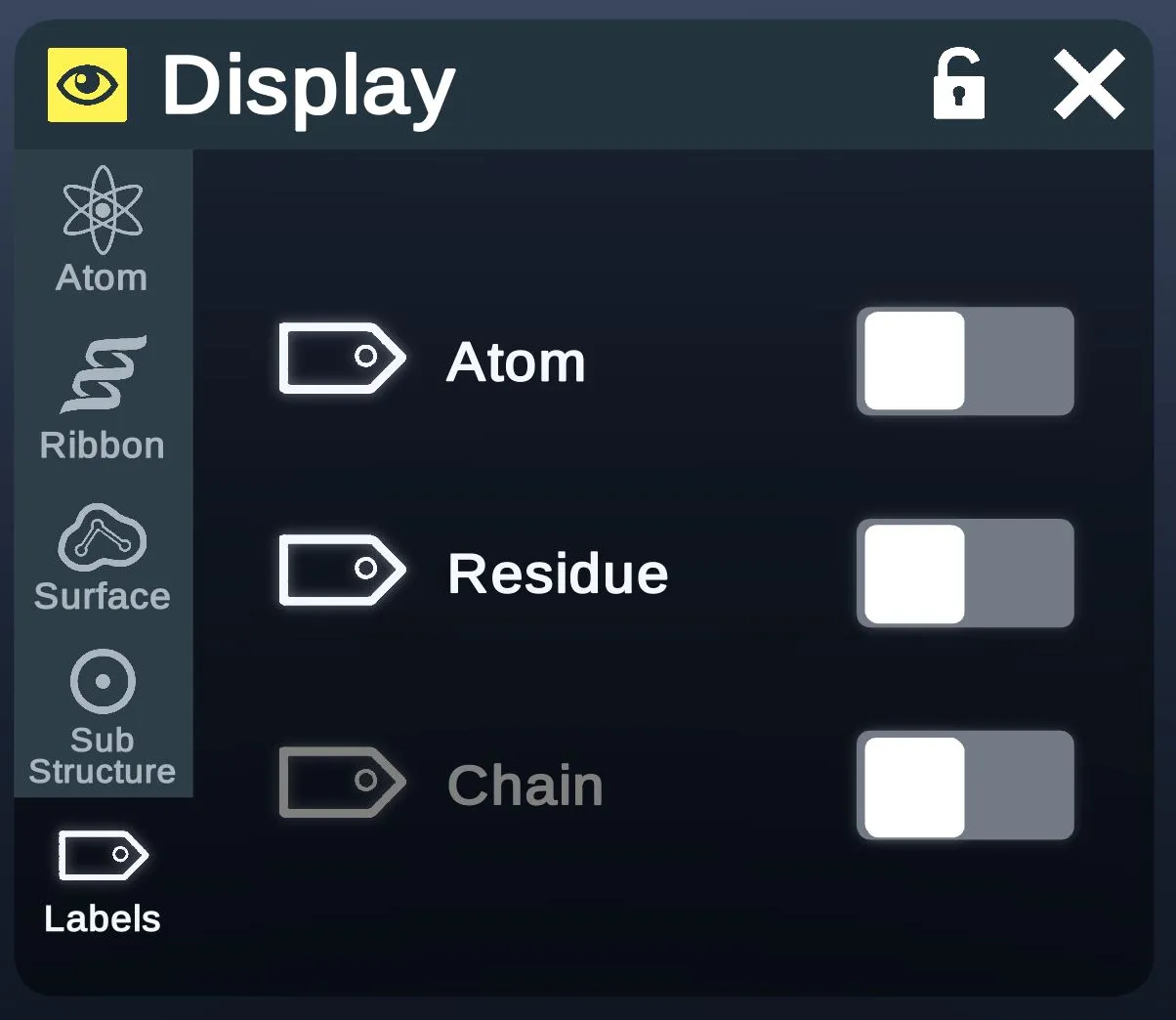
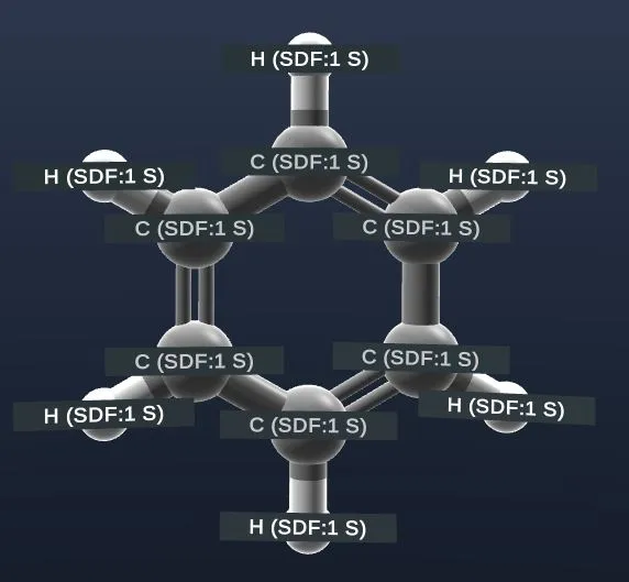
- AtomShow labels on atoms.
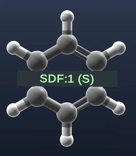
- ResidueShow labels on residues.
- ChainShow labels on chains. Grayed out in this screenshot.
| Atom | Residue |
|---|---|
 |  |
Color
The Color menu applies a solid color or a color scheme to the selected structures.
Picker
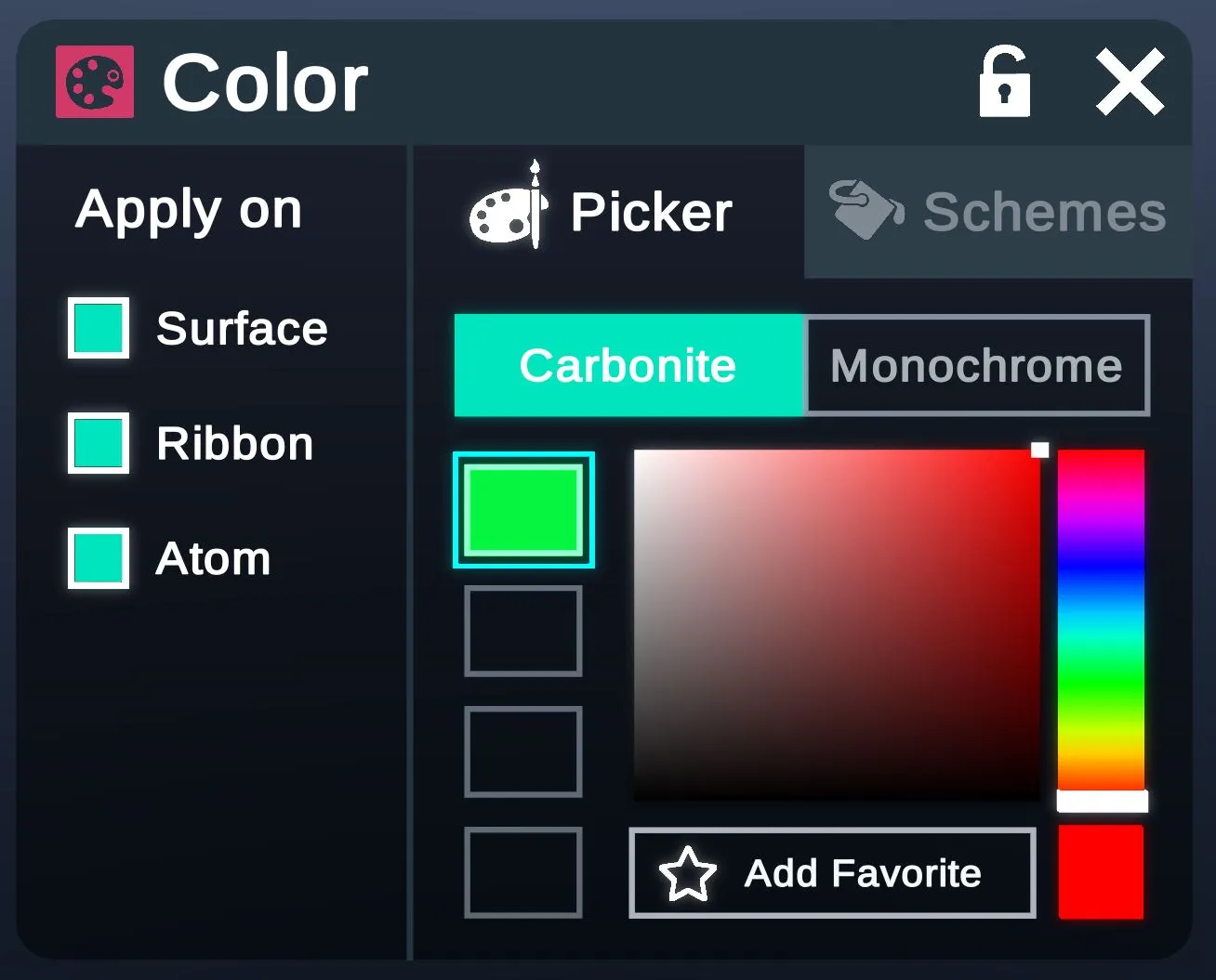
Colors from the picker apply as one solid color.
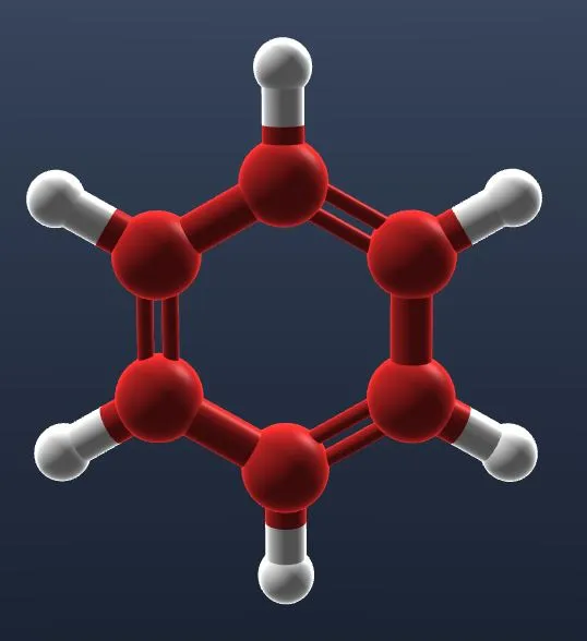
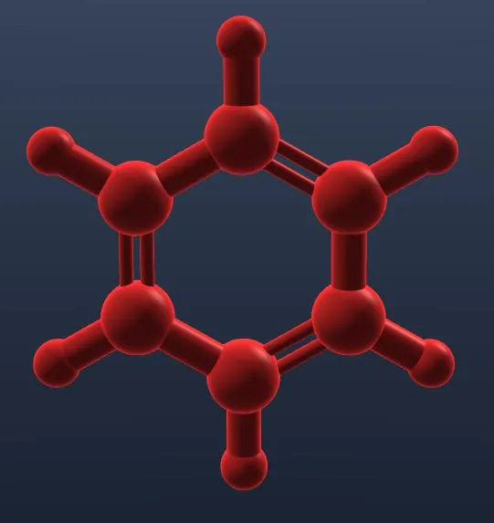
- Carbonite color applies only to carbons.
- Monochrome color applies to every atom.
- Color pickerPicks a color to apply in a solid style.
- Add FavoriteAdds the picked color as a favorite.
- Apply onWhich parts get the color: Surface, Ribbon and Atom.
| Carbonite | Monochrome |
|---|---|
 |  |
Schemes
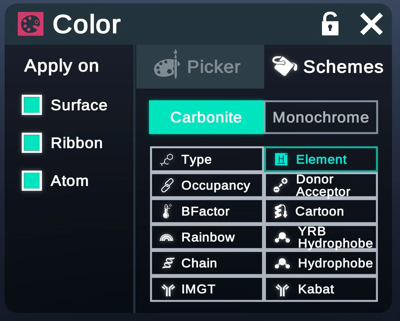
A scheme colors the selected structures by a property, such as element or chain.
- As in the Picker tab.
- Colors each residue by its type.
- Colors atoms by element. Picked here.
- Colors the selected structures by occupancy.
- Colors hydrogen-bond donors and acceptors.
- Colors the selected structures by b-factor.
- Colors by secondary structure.
- Colors the selected structures in rainbow colors.
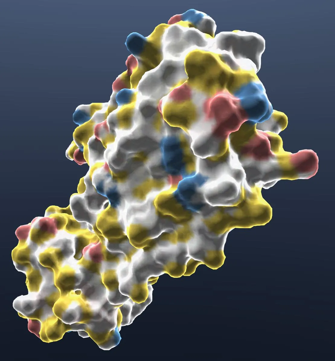
- Colors carbons not bound to nitrogen or oxygen yellow, the charged oxygens of Asp and Glu red, and the charged nitrogens of Lys and Arg blue. Everything else is white.
- Gives each chain its own color.
- Colors by hydrophobicity.
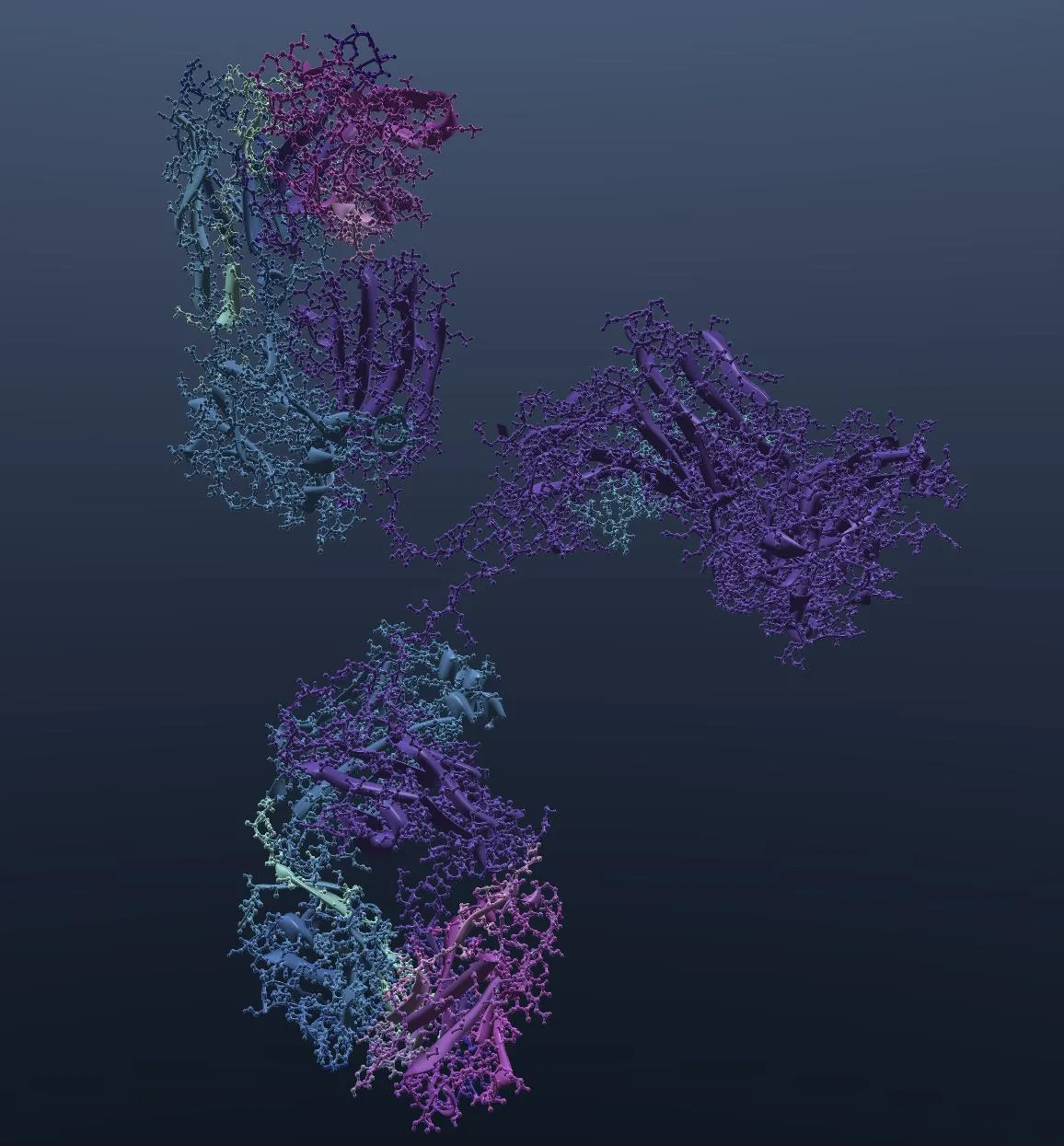
- Colors the selected antibody structures by their regions.
- Colors antibody regions as the Kabat numbering scheme defines them.
| YRB Hydrophobe | IMGT |
|---|---|
 |  |
Modify
Edit
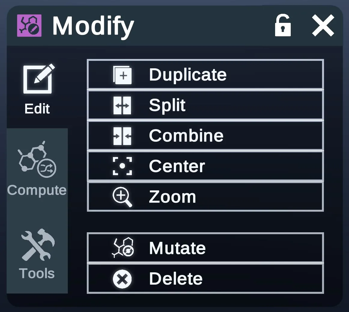
The Edit tab's buttons change the selected structures.
- DuplicateCopies the selected structures.
- Splits the selected structures.
- Combines the selected structures.
- Centers the selected structures.
- ZoomZooms to the selected structures.
- Opens the Mutate menu.
- Deletes the selected structures.
Compute
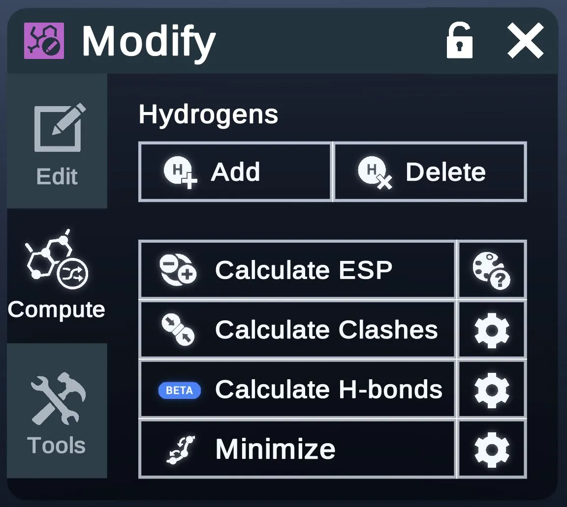
The Compute tab runs calculations on the selected structures, such as clashes and h-bonds. It can also add hydrogens to them and minimize them.
- Under Hydrogens: adds hydrogens to the selected structures.
- Under Hydrogens: deletes hydrogens from the selected structures.
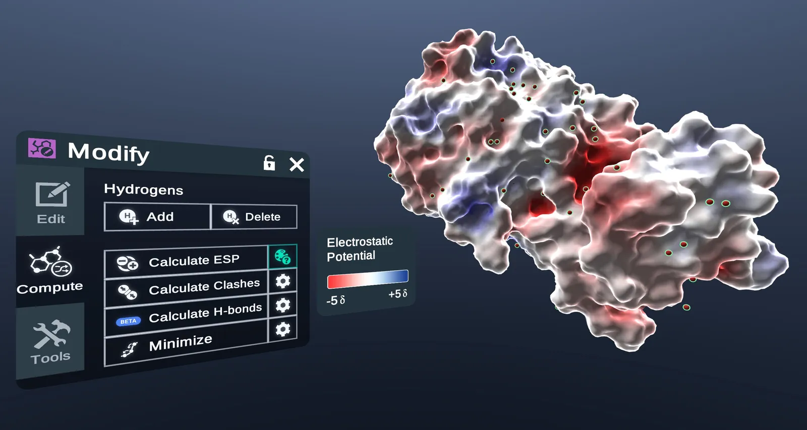
- Calculates the electrostatic potential (ESP) of the selected structure.
- ESP color keyShows the Electrostatic Potential color key. It's on in the picture below.
- Calculates atom clashes.
- Calculates h-bonds. Marked BETA.
- Minimizes the potential energy of the selected structures.
- SettingsOpens the Clashes, H-bonds or Minimize menu, shown below.
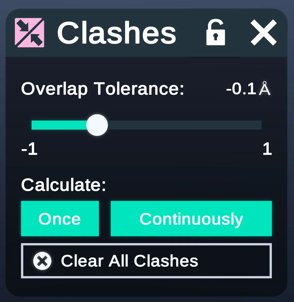
Calculate Clashes: sets the clash tolerance.
- The tolerance, from -1 to 1 Å.
- Calculates clashes one time, or keeps calculating them.
- Clears every clash.
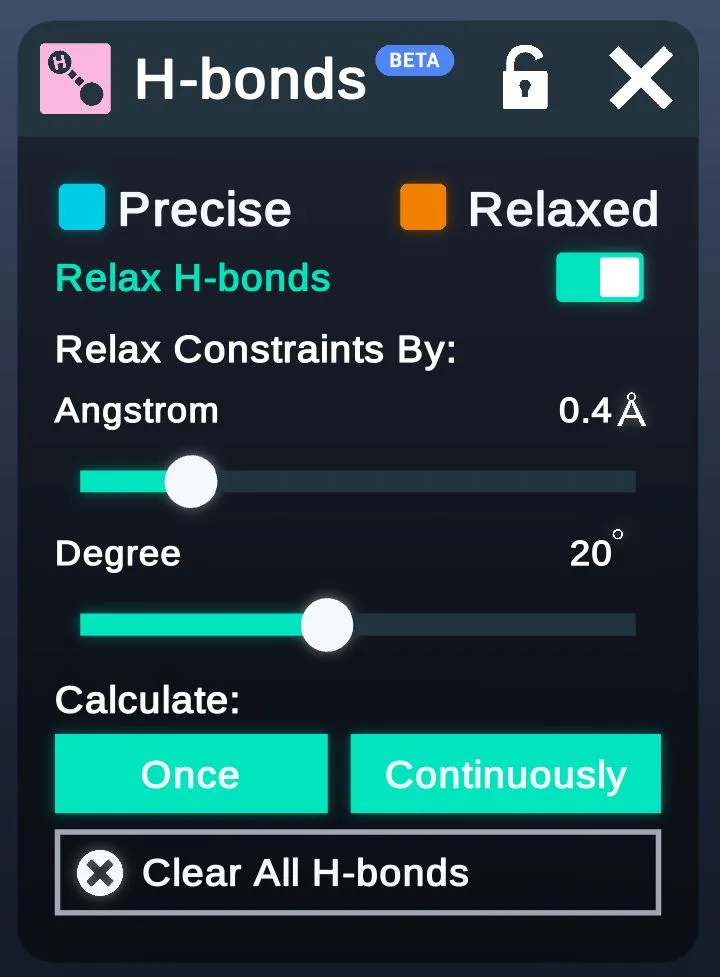
Calculate H-bonds: sets the threshold for finding H-bonds.
- The color key: cyan for precise h-bonds, orange for relaxed ones.
- Relaxes the h-bond constraints. On here.
- The threshold, in Angstrom and degrees.
- Calculates h-bonds one time, or keeps calculating them.
- Clears every h-bond.
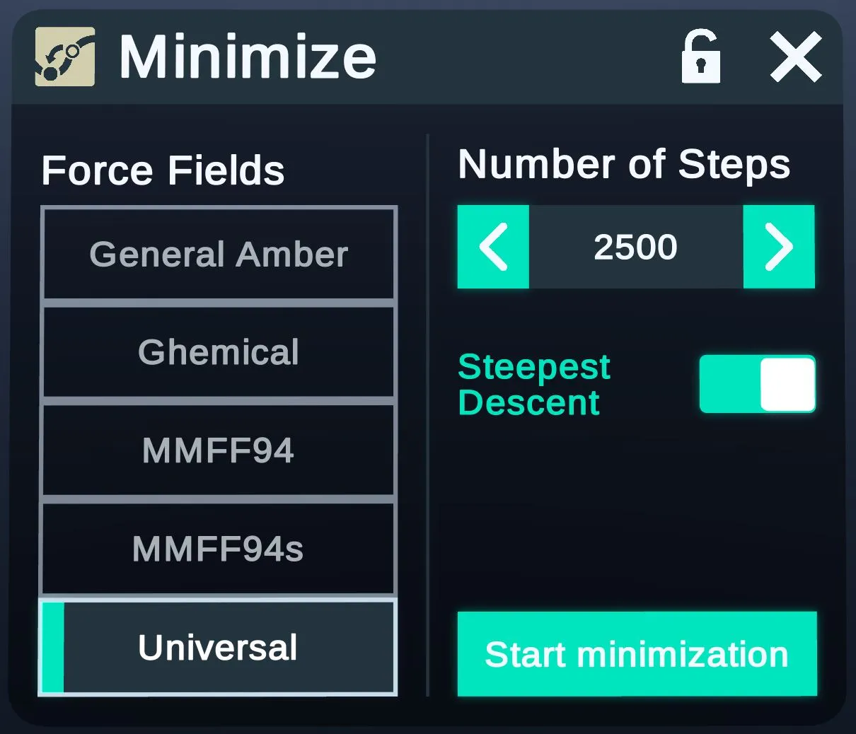
Minimize: sets the force field and steps used to minimize the selected structures.
- The force field to use.
- The arrows change the number of steps.
- Uses the steepest descent method for the minimization. On here.
- Starts minimizing the selected structures.
Tools
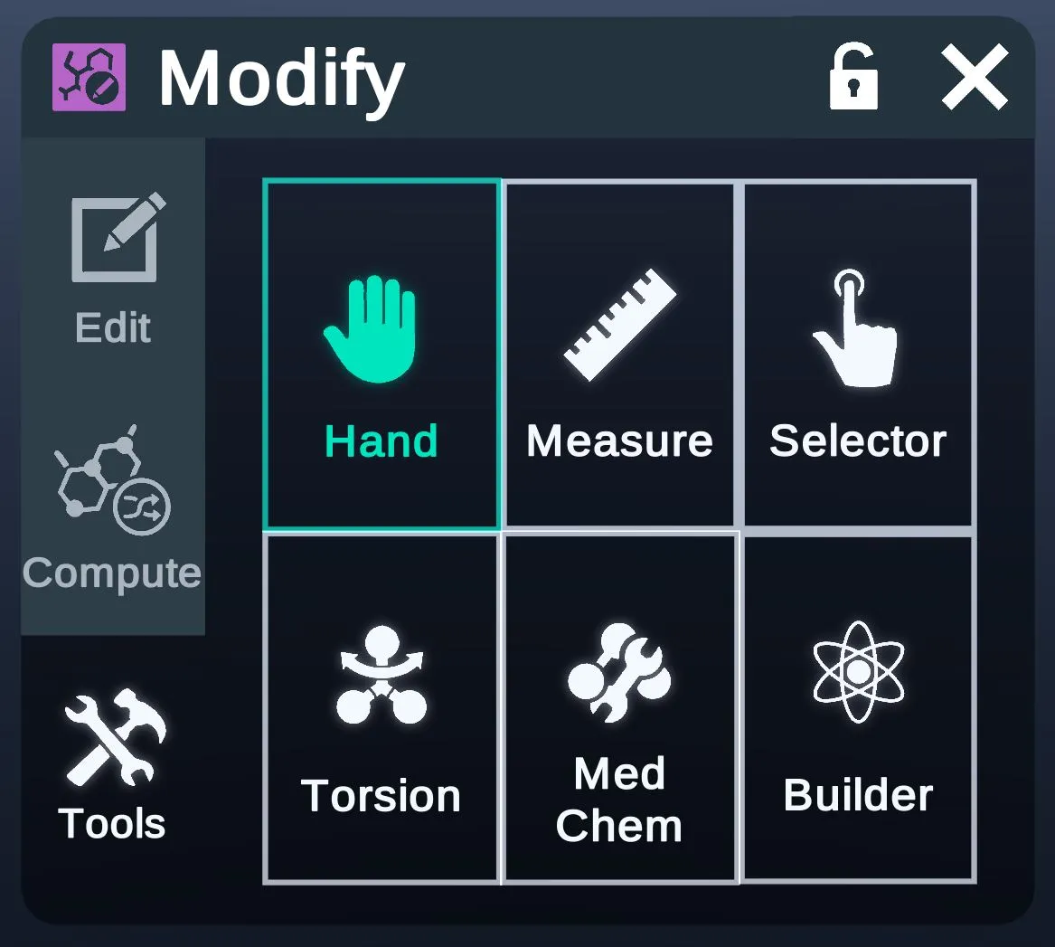
The Tools tab switches the tool in your hand, to measure, select or edit structures directly.
Mutate
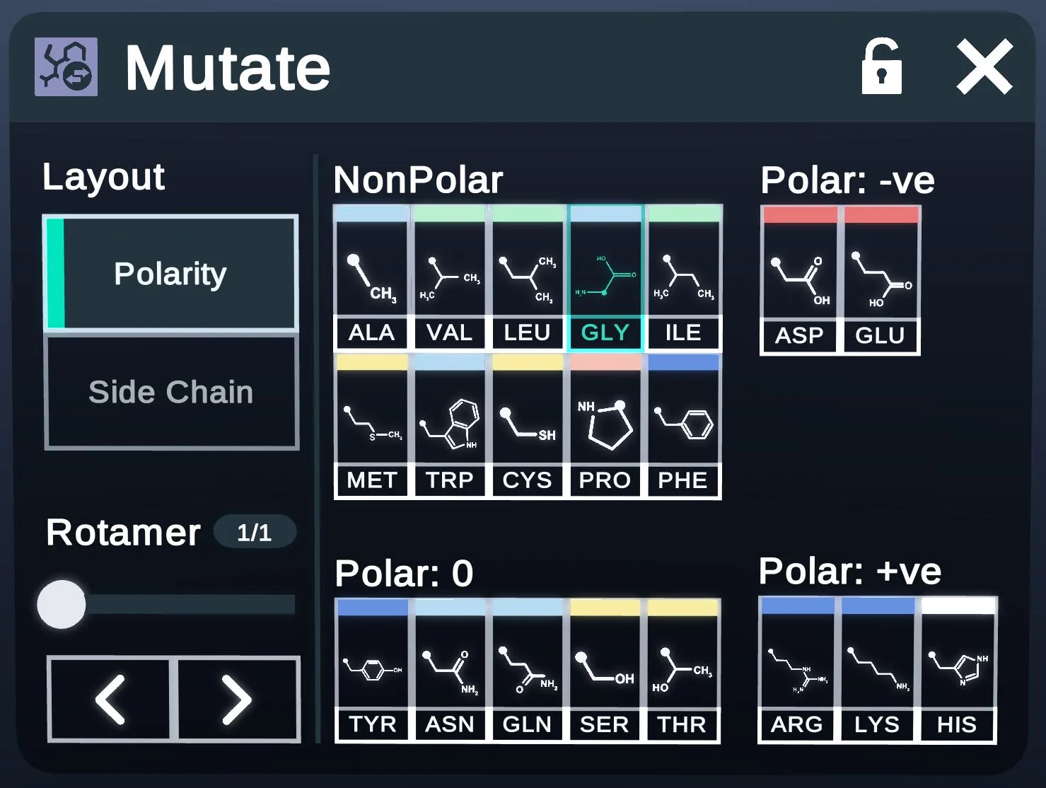
The Mutate menu swaps the R-groups of the selected amino acids.
- Under Layout: lays the amino acids out by polarity, as here.
- Under Layout: lays them out by side chain.
- Nonpolar amino acids.
- Polar, negatively charged.
- Polar, uncharged.
- Polar, positively charged.
- The slider and arrows step through rotamers.
Select
The Selection menu changes the selection by atom type, residue type or distance.
Refine
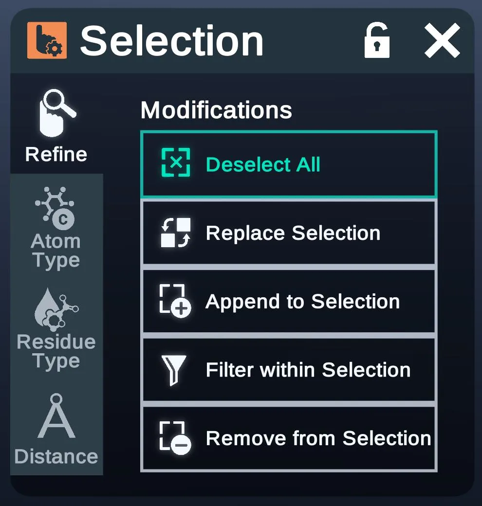
The Refine tab clears the selection, or replaces, adds to, filters or removes from it.
- Deselect AllDeselects everything.
- Replace SelectionOpens the Replace Selection menu, shown below.
- Append to SelectionAdds to the current selection.
- Filter within SelectionFilters the current selection.
- Remove from SelectionRemoves from the current selection.
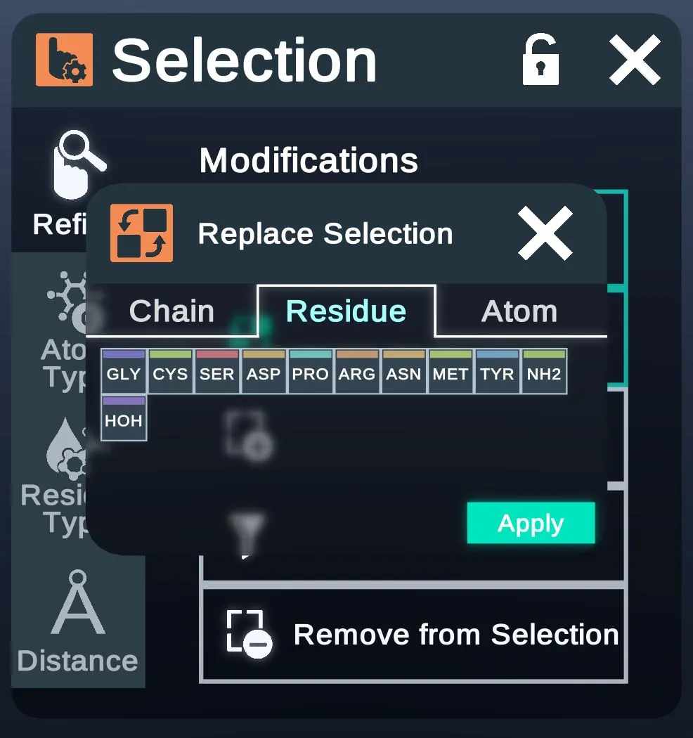
Replace Selection
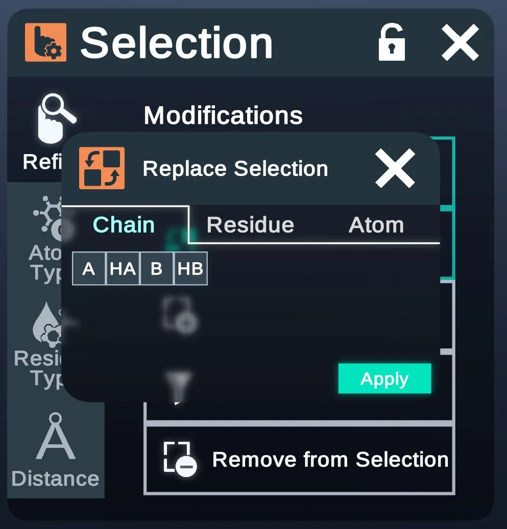
- ChainLists the chains, such as A, HA, B and HB.
- Lists the residue names, as here.
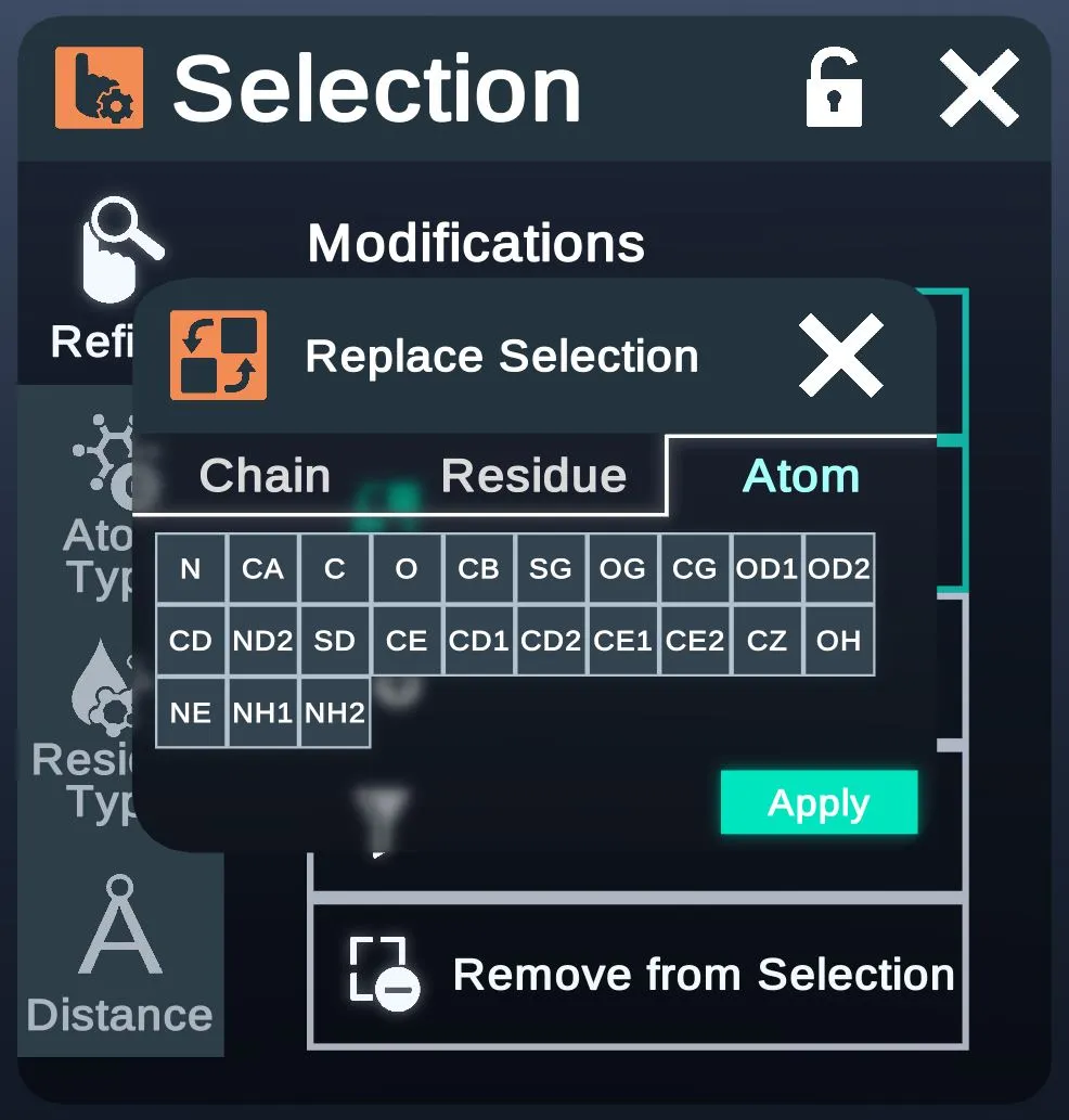
- AtomLists the atom names, such as N, CA and CB.
- ListThe items on the open tab.
- ApplyReplaces the selection.
| Chain tab | Atom tab |
|---|---|
 |  |
Atom Type
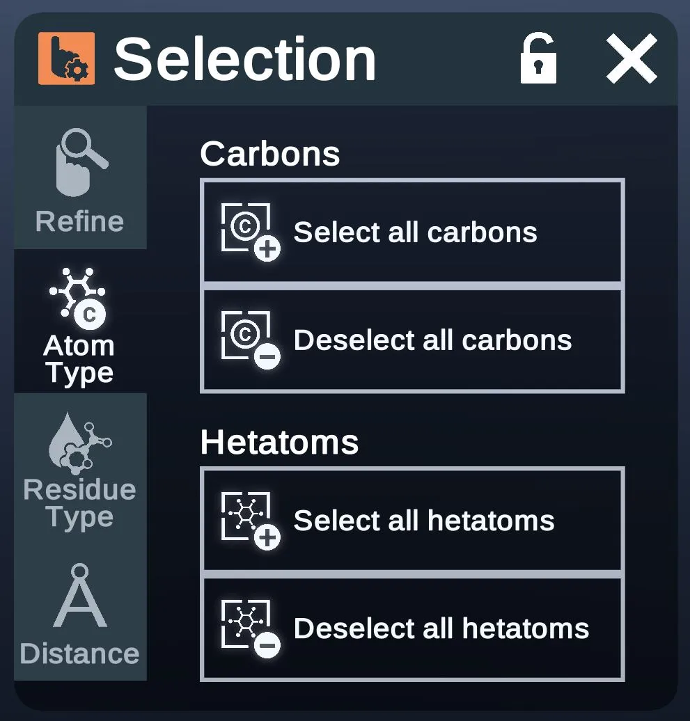
The Atom Type tab selects or deselects every carbon or every hetatom.
- Select all carbonsSelects every carbon atom.
- Deselect all carbonsDeselects every carbon atom.
- Select all hetatomsSelects every hetatom.
- Deselect all hetatomsDeselects every hetatom.
Residue Type
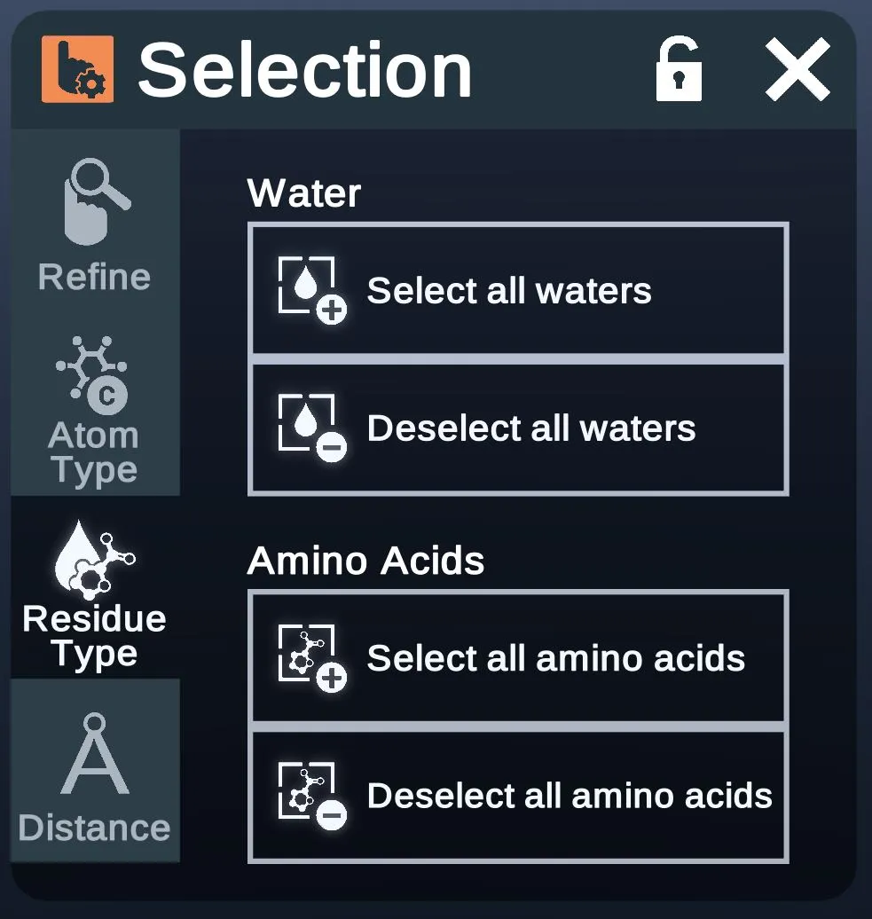
The Residue Type tab selects or deselects every water or every amino acid.
- Select all watersSelects every water.
- Deselect all watersDeselects every water.
- Select all amino acidsSelects every amino acid.
- Deselect all amino acidsDeselects every amino acid.
Distance
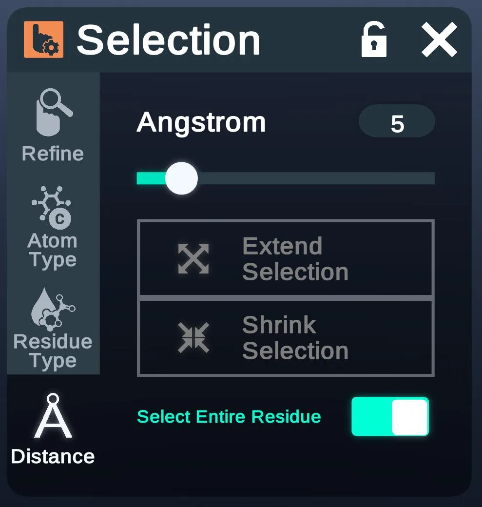
The Distance tab grows or shrinks the selection by a distance in Å.
- AngstromThe distance, in Angstroms. 5 here.
- Extend SelectionGrows the selection by the distance above. Grayed out here.
- Shrink SelectionShrinks the selection by the distance above. Grayed out here.
- When on, distance selection takes whole residues.
Macro
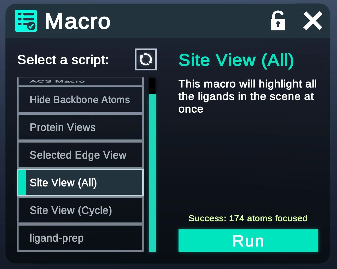
The macros page has more.
Content Discovery
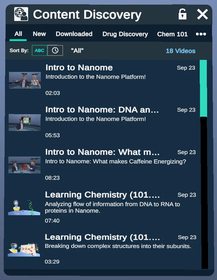
Tutorials of advanced concepts and features in Nanome, presented using spatial recordings.
Advanced Tutorials
In version 1.24.6, Content Discovery replaced the Advanced Tutorials menu.
Spatial Recordings
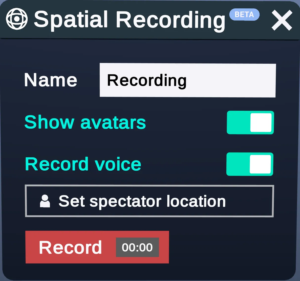
A spatial recording captures your entire workspace, including structures, menus and virtual movements, to play back later. During playback you can pause and even interact with the structures in the recording.
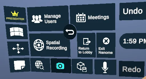
Spatial recordings can be accessed from either the Wrist Menu (see below) or the Action Menu.
Spatial recordings were a beta feature in Nanome 1.20. Recordings made in 1.20 can't be played in later versions.